4BXW | pdb_00004bxw

Crystal Structure of the Prothrombinase Complex from the Venom of Pseudonaja Textilis


Experimental Data Snapshot

  • Method: X-RAY DIFFRACTION
  • Resolution: 2.71 Å
  • R-Value Free: 
    0.233 (Depositor), 0.216 (DCC) 
  • R-Value Work: 
    0.155 (Depositor), 0.156 (DCC) 
  • R-Value Observed: 
    0.159 (Depositor) 

Starting Model: experimental
View more details

wwPDB Validation 3D Report Full Report

Validation slider image for 4BXW

Ligand Structure Quality Assessment 


This is version 1.3 of the entry. See complete history

Literature

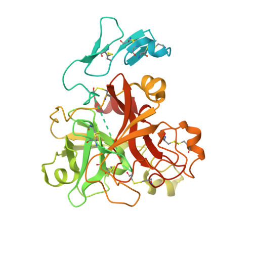
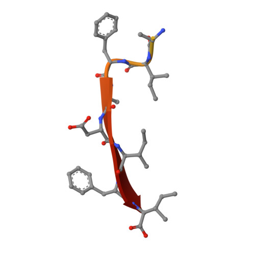
Crystal Structure of the Prothrombinase Complex from the Venom of Pseudonaja Textilis.

Lechtenberg, B.C.Murray-Rust, T.A.Johnson, D.J.Adams, T.E.Krishnaswamy, S.Camire, R.M.Huntington, J.A.

(2013) Blood 122: 2777

  • DOI: https://doi.org/10.1182/blood-2013-06-511733
  • Primary Citation Related Structures: 
    4BXS, 4BXW

  • PubMed Abstract: 

    The prothrombinase complex, composed of the protease factor (f)Xa and cofactor fVa, efficiently converts prothrombin to thrombin by specific sequential cleavage at 2 sites. How the complex assembles and its mechanism of prothrombin processing are of central importance to human health and disease, because insufficient thrombin generation is the root cause of hemophilia, and excessive thrombin production results in thrombosis. Efforts to determine the crystal structure of the prothrombinase complex have been thwarted by the dependence of complex formation on phospholipid membrane association. Pseutarin C is an intrinsically stable prothrombinase complex preassembled in the venom gland of the Australian Eastern Brown Snake (Pseudonaja textilis). Here we report the crystal structures of the fX-fV complex and of activated fXa from P textilis venom and the derived model of active pseutarin C. Structural analysis supports a single substrate binding channel on fVa, to which prothrombin and the intermediate meizothrombin bind in 2 different orientations, providing insight into the architecture and mechanism of the prothrombinase complex-the molecular engine of blood coagulation.


  • Organizational Affiliation
    • Department of Haematology, Cambridge Institute for Medical Research, University of Cambridge, Cambridge, United Kingdom; and.

Macromolecule Content 

  • Total Structure Weight: 97.23 kDa 
  • Atom Count: 4,538 
  • Modeled Residue Count: 581 
  • Deposited Residue Count: 864 
  • Unique protein chains: 2

Macromolecules

Find similar proteins by:|  3D Structure
Entity ID: 1
MoleculeChains  Sequence LengthOrganismDetailsImage
FACTOR XA
A, B
423Pseudonaja textilisMutation(s): 0 
EC: 3.4.21.6
UniProt
Find proteins for Q56VR3 (Pseudonaja textilis)
Explore Q56VR3 
Go to UniProtKB:  Q56VR3
Entity Groups
Sequence Clusters30% Identity50% Identity70% Identity90% Identity95% Identity100% Identity
UniProt GroupQ56VR3
Sequence Annotations
Expand
Reference Sequence
Find similar proteins by:  Sequence   |   3D Structure  
Entity ID: 2
MoleculeChains  Sequence LengthOrganismDetailsImage
COAGULATION FACTOR VC [auth F]18Escherichia coliMutation(s): 0 
UniProt
Find proteins for Q593B6 (Pseudonaja textilis)
Explore Q593B6 
Go to UniProtKB:  Q593B6
Entity Groups
Sequence Clusters30% Identity50% Identity70% Identity90% Identity95% Identity100% Identity
UniProt GroupQ593B6
Sequence Annotations
Expand
Reference Sequence

Small Molecules

Ligands 3 Unique
IDChains Name / Formula / InChI Key2D Diagram3D Interactions
0GJ

Query on 0GJ


Download:Ideal Coordinates CCD File
D [auth A],
G [auth B]
L-alpha-glutamyl-N-{(1S)-4-{[amino(iminio)methyl]amino}-1-[(1S)-2-chloro-1-hydroxyethyl]butyl}glycinamide
C14 H28 Cl N6 O5
XELWNHKFCNMWQO-LPEHRKFASA-O
GOL

Query on GOL


Download:Ideal Coordinates CCD File
F [auth A],
H [auth B],
I [auth B]
GLYCEROL
C3 H8 O3
PEDCQBHIVMGVHV-UHFFFAOYSA-N
NA

Query on NA


Download:Ideal Coordinates CCD File
E [auth A]SODIUM ION
Na
FKNQFGJONOIPTF-UHFFFAOYSA-N

Biologically Interesting Molecules (External Reference) 

1 Unique

Experimental Data & Validation

Experimental Data

  • Method: X-RAY DIFFRACTION
  • Resolution: 2.71 Å
  • R-Value Free:  0.233 (Depositor), 0.216 (DCC) 
  • R-Value Work:  0.155 (Depositor), 0.156 (DCC) 
  • R-Value Observed: 0.159 (Depositor) 
Space Group: I 21 3
Unit Cell:
Length ( Å )Angle ( ˚ )
a = 179.31α = 90
b = 179.31β = 90
c = 179.31γ = 90
Software Package:
Software NamePurpose
REFMACrefinement
MOSFLMdata reduction
Aimlessdata scaling
PHASERphasing

Structure Validation

View Full Validation Report


Ligand Structure Quality Assessment 


Entry History 

Deposition Data

Revision History  (Full details and data files)

  • Version 1.0: 2013-07-31
    Type: Initial release
  • Version 1.1: 2013-10-30
    Changes: Database references
  • Version 1.2: 2023-12-20
    Changes: Data collection, Database references, Derived calculations, Other, Refinement description
  • Version 1.3: 2024-11-20
    Changes: Structure summary