Influence of the Agrc-Agra Complex in the Response Time of Staphylococcus Aureus Quorum Sensing
Srivastava, S.K., Rajasree, K., Fasim, A., Arakere, G., Gopal, B.(2014) J Bacteriol 196: 2876
- PubMed: 24858185
- DOI: https://doi.org/10.1128/JB.01530-14
- Primary Citation of Related Structures:
4BXI - PubMed Abstract:
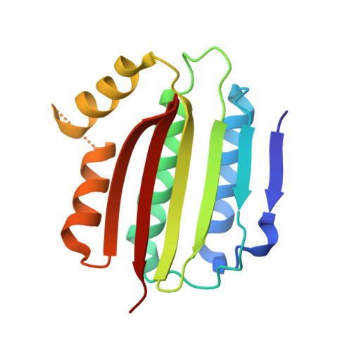
The Staphylococcus aureus agr quorum-sensing system plays a major role in the transition from the persistent to the virulent phenotype. S. aureus agr type I to IV strains are characterized by mutations in the sensor domain of the histidine kinase AgrC and differences in the sequences of the secreted autoinducing peptides (AIP). Here we demonstrate that interactions between the cytosolic domain of AgrC (AgrCCyto) and the response regulator domain of AgrA (AgrARR) dictate the spontaneity of the cellular response to AIP stimuli. The crystal structure of AgrCCyto provided a basis for a mechanistic model of AgrC-AgrA interactions. This model enabled an analysis of the biochemical and biophysical parameters of AgrC-AgrA interactions in the context of the conformational features of the AgrC-AgrA complex. This analysis revealed distinct sequence and conformational features that determine the affinity, specificity, and kinetics of the phosphotransfer reaction. This step, which governs the response time for transcriptional reengineering triggered by an AIP stimulus, is independent of the agr type and similar for agonist and antagonist stimuli. These experimental data could serve as a basis on which to validate simulations of the quorum-sensing response and for strategies that employ the agr quorum-sensing system to combat biofilm formation in S. aureus infections.
Organizational Affiliation:
Molecular Biophysics Unit, Indian Institute of Science, Bangalore, India.


