4BVG | pdb_00004bvg

CRYSTAL STRUCTURE OF HUMAN SIRT3 IN COMPLEX WITH NATIVE ALKYLIMIDATE FORMED FROM ACETYL-LYSINE ACS2-PEPTIDE CRYSTALLIZED IN PRESENCE OF THE INHIBITOR EX-527


Experimental Data Snapshot

  • Method: X-RAY DIFFRACTION
  • Resolution: 2.50 Å
  • R-Value Free: 
    0.248 (Depositor), 0.250 (DCC) 
  • R-Value Work: 
    0.182 (Depositor), 0.180 (DCC) 
  • R-Value Observed: 
    0.186 (Depositor) 

Starting Model: experimental
View more details

wwPDB Validation   3D Report Full Report


Ligand Structure Quality Assessment 

Created with Raphaël 2.3.0Worse 01 BetterLigand structure goodness of fit to experimental dataBest fitted XYQClick on this verticalbar to view details

This is version 2.2 of the entry. See complete history


Literature

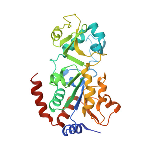
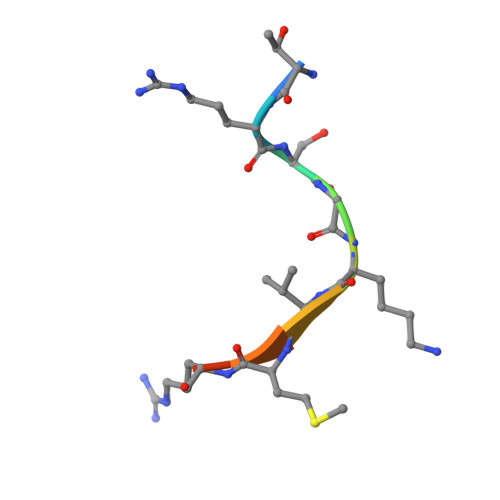
Ex-527 inhibits Sirtuins by exploiting their unique NAD+-dependent deacetylation mechanism.

Gertz, M.Fischer, F.Nguyen, G.T.Lakshminarasimhan, M.Schutkowski, M.Weyand, M.Steegborn, C.

(2013) Proc Natl Acad Sci U S A 110: E2772-E2781

  • DOI: https://doi.org/10.1073/pnas.1303628110
  • Primary Citation of Related Structures:  
    4BUZ, 4BV2, 4BV3, 4BVB, 4BVE, 4BVF, 4BVG, 4BVH

  • PubMed Abstract: 

    Sirtuins are protein deacetylases regulating metabolism and stress responses. The seven human Sirtuins (Sirt1-7) are attractive drug targets, but Sirtuin inhibition mechanisms are mostly unidentified. We report the molecular mechanism of Sirtuin inhibition by 6-chloro-2,3,4,9-tetrahydro-1H-carbazole-1-carboxamide (Ex-527). Inhibitor binding to potently inhibited Sirt1 and Thermotoga maritima Sir2 and to moderately inhibited Sirt3 requires NAD(+), alone or together with acetylpeptide. Crystal structures of several Sirtuin inhibitor complexes show that Ex-527 occupies the nicotinamide site and a neighboring pocket and contacts the ribose of NAD(+) or of the coproduct 2'-O-acetyl-ADP ribose. Complex structures with native alkylimidate and thio-analog support its catalytic relevance and show, together with biochemical assays, that only the coproduct complex is relevant for inhibition by Ex-527, which stabilizes the closed enzyme conformation preventing product release. Ex-527 inhibition thus exploits Sirtuin catalysis, and kinetic isoform differences explain its selectivity. Our results provide insights in Sirtuin catalysis and inhibition with important implications for drug development.


  • Organizational Affiliation

    Department of Biochemistry and Research Center for Bio-Macromolecules, University of Bayreuth, 95440 Bayreuth, Germany.


Macromolecules
Find similar proteins by:  (by identity cutoff)  |  3D Structure
Entity ID: 1
MoleculeChains Sequence LengthOrganismDetailsImage
NAD-DEPENDENT PROTEIN DEACETYLASE SIRTUIN-3, MITOCHONDRIAL284Homo sapiensMutation(s): 0 
EC: 3.5.1 (PDB Primary Data), 2.3.1 (UniProt), 2.3.1.286 (UniProt)
UniProt & NIH Common Fund Data Resources
Find proteins for Q9NTG7 (Homo sapiens)
Explore Q9NTG7 
Go to UniProtKB:  Q9NTG7
PHAROS:  Q9NTG7
GTEx:  ENSG00000142082 
Entity Groups  
Sequence Clusters30% Identity50% Identity70% Identity90% Identity95% Identity100% Identity
UniProt GroupQ9NTG7
Sequence Annotations
Expand
  • Reference Sequence

Find similar proteins by:  Sequence   |   3D Structure  

Entity ID: 2
MoleculeChains Sequence LengthOrganismDetailsImage
ACETYL-COENZYME A SYNTHETASE 2-LIKE, MITOCHONDRIAL10Homo sapiensMutation(s): 0 
EC: 6.2.1.1 (PDB Primary Data), 6.2.1.17 (UniProt)
UniProt & NIH Common Fund Data Resources
Find proteins for Q9NUB1 (Homo sapiens)
Explore Q9NUB1 
Go to UniProtKB:  Q9NUB1
PHAROS:  Q9NUB1
GTEx:  ENSG00000154930 
Entity Groups  
Sequence Clusters30% Identity50% Identity70% Identity90% Identity95% Identity100% Identity
UniProt GroupQ9NUB1
Sequence Annotations
Expand
  • Reference Sequence
Small Molecules
Ligands 6 Unique
IDChains Name / Formula / InChI Key2D Diagram3D Interactions
XYQ
Query on XYQ

Download Ideal Coordinates CCD File 
Q [auth B](2R,3R,4S,5R)-5-({[(R)-{[(R)-{[(2R,3S,4R,5R)-5-(6-AMINO-9H-PURIN-9-YL)-3,4-DIHYDROXYTETRAHYDROFURAN-2-YL]METHOXY}(HYDROXY)PHOSPHORYL]OXY}(HYDROXY)PHOSPHORYL]OXY}METHYL)-3,4-DIHYDROXYTETRAHYDROFURAN-2-YL ACETATE
C17 H25 N5 O15 P2
IJOUKWCBVUMMCR-DLFWLGJNSA-N
PEG
Query on PEG

Download Ideal Coordinates CCD File 
O [auth A]DI(HYDROXYETHYL)ETHER
C4 H10 O3
MTHSVFCYNBDYFN-UHFFFAOYSA-N
SO4
Query on SO4

Download Ideal Coordinates CCD File 
L [auth A],
M [auth A]
SULFATE ION
O4 S
QAOWNCQODCNURD-UHFFFAOYSA-L
GOL
Query on GOL

Download Ideal Coordinates CCD File 
G [auth A],
H [auth A],
I [auth A]
GLYCEROL
C3 H8 O3
PEDCQBHIVMGVHV-UHFFFAOYSA-N
ZN
Query on ZN

Download Ideal Coordinates CCD File 
P [auth A]ZINC ION
Zn
PTFCDOFLOPIGGS-UHFFFAOYSA-N
EDO
Query on EDO

Download Ideal Coordinates CCD File 
C [auth A]
D [auth A]
E [auth A]
F [auth A]
J [auth A]
1,2-ETHANEDIOL
C2 H6 O2
LYCAIKOWRPUZTN-UHFFFAOYSA-N
Experimental Data & Validation

Experimental Data

  • Method: X-RAY DIFFRACTION
  • Resolution: 2.50 Å
  • R-Value Free:  0.248 (Depositor), 0.250 (DCC) 
  • R-Value Work:  0.182 (Depositor), 0.180 (DCC) 
  • R-Value Observed: 0.186 (Depositor) 
Space Group: C 2 2 21
Unit Cell:
Length ( Å )Angle ( ˚ )
a = 78.023α = 90
b = 131.329β = 90
c = 76.508γ = 90
Software Package:
Software NamePurpose
REFMACrefinement
XDSdata reduction
XSCALEdata scaling
PHASERphasing

Structure Validation

View Full Validation Report


Ligand Structure Quality Assessment 

Created with Raphaël 2.3.0Worse 01 BetterLigand structure goodness of fit to experimental dataBest fitted XYQClick on this verticalbar to view details

Entry History 

Deposition Data

Revision History  (Full details and data files)

  • Version 1.0: 2013-07-17
    Type: Initial release
  • Version 1.1: 2013-08-07
    Changes: Database references
  • Version 2.0: 2019-01-30
    Changes: Atomic model, Data collection, Database references, Derived calculations, Experimental preparation, Non-polymer description, Structure summary
  • Version 2.1: 2023-12-20
    Changes: Data collection, Database references, Derived calculations, Other, Refinement description
  • Version 2.2: 2024-10-16
    Changes: Structure summary