4BRU | pdb_00004bru

Crystal structure of the yeast Dhh1-Edc3 complex


Experimental Data Snapshot

  • Method: X-RAY DIFFRACTION
  • Resolution: 3.25 Å
  • R-Value Free: 
    0.246 (Depositor), 0.274 (DCC) 
  • R-Value Work: 
    0.208 (Depositor), 0.245 (DCC) 
  • R-Value Observed: 
    0.210 (Depositor) 

Starting Model: experimental
View more details

wwPDB Validation 3D Report Full Report

Validation slider image for 4BRU

This is version 1.2 of the entry. See complete history

Literature

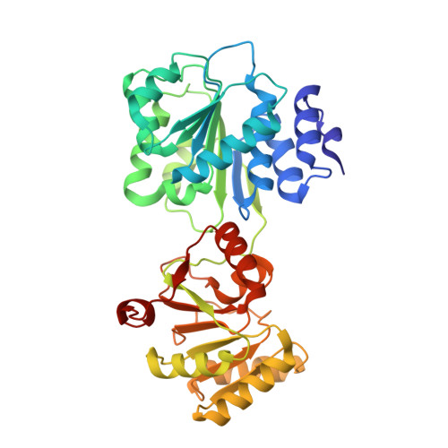
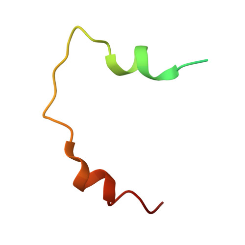
Structural Analysis of the Yeast Dhh1-Pat1 Complex Reveals How Dhh1 Engages Pat1, Edc3 and RNA in Mutually Exclusive Interactions

Sharif, H.Ozgur, S.Sharma, K.Basquin, C.Urlaub, H.Conti, E.

(2013) Nucleic Acids Res 41: 8377

  • DOI: https://doi.org/10.1093/nar/gkt600
  • Primary Citation Related Structures: 
    4BRU, 4BRW

  • PubMed Abstract: 

    Translational repression and deadenylation of eukaryotic mRNAs result either in the sequestration of the transcripts in a nontranslatable pool or in their degradation. Removal of the 5' cap structure is a crucial step that commits deadenylated mRNAs to 5'-to-3' degradation. Pat1, Edc3 and the DEAD-box protein Dhh1 are evolutionary conserved factors known to participate in both translational repression and decapping, but their interplay is currently unclear. We report the 2.8 Å resolution structure of yeast Dhh1 bound to the N-terminal domain of Pat1. The structure shows how Pat1 wraps around the C-terminal RecA domain of Dhh1, docking onto the Phe-Asp-Phe (FDF) binding site. The FDF-binding site of Dhh1 also recognizes Edc3, revealing why the binding of Pat1 and Edc3 on Dhh1 are mutually exclusive events. Using co-immunoprecipitation assays and structure-based mutants, we demonstrate that the mode of Dhh1-Pat1 recognition is conserved in humans. Pat1 and Edc3 also interfere and compete with the RNA-binding properties of Dhh1. Mapping the RNA-binding sites on Dhh1 with a crosslinking-mass spectrometry approach shows a large RNA-binding surface around the C-terminal RecA domain, including the FDF-binding pocket. The results suggest a model for how Dhh1-containing messenger ribonucleoprotein particles might be remodeled upon Pat1 and Edc3 binding.


  • Organizational Affiliation
    • Structural Cell Biology Department, Max Planck Institute of Biochemistry, Am Klopferspitz 18, Martinsried/Munich, D-82152 Germany and Cellular Biochemistry Department, Max Planck Institute of Biophysical Chemistry, Am Faßberg 11, 37077 Göttingen, Germany.

Macromolecule Content 

  • Total Structure Weight: 48.17 kDa 
  • Atom Count: 3,110 
  • Modeled Residue Count: 404 
  • Deposited Residue Count: 420 
  • Unique protein chains: 2

Macromolecules

Find similar proteins by:|  3D Structure
Entity ID: 1
MoleculeChains  Sequence LengthOrganismDetailsImage
ATP-DEPENDENT RNA HELICASE DHH1377Saccharomyces cerevisiae S288CMutation(s): 2 
EC: 3.6.4.13
UniProt
Find proteins for P39517 (Saccharomyces cerevisiae (strain ATCC 204508 / S288c))
Explore P39517 
Go to UniProtKB:  P39517
Entity Groups
Sequence Clusters30% Identity50% Identity70% Identity90% Identity95% Identity100% Identity
UniProt GroupP39517
Sequence Annotations
Expand
Reference Sequence
Find similar proteins by:|  3D Structure
Entity ID: 2
MoleculeChains  Sequence LengthOrganismDetailsImage
ENHANCER OF MRNA-DECAPPING PROTEIN 343Saccharomyces cerevisiae S288CMutation(s): 0 
UniProt
Find proteins for P39998 (Saccharomyces cerevisiae (strain ATCC 204508 / S288c))
Explore P39998 
Go to UniProtKB:  P39998
Entity Groups
Sequence Clusters30% Identity50% Identity70% Identity90% Identity95% Identity100% Identity
UniProt GroupP39998
Sequence Annotations
Expand
Reference Sequence

Experimental Data & Validation

Experimental Data

  • Method: X-RAY DIFFRACTION
  • Resolution: 3.25 Å
  • R-Value Free:  0.246 (Depositor), 0.274 (DCC) 
  • R-Value Work:  0.208 (Depositor), 0.245 (DCC) 
  • R-Value Observed: 0.210 (Depositor) 
Space Group: P 41 2 2
Unit Cell:
Length ( Å )Angle ( ˚ )
a = 105.831α = 90
b = 105.831β = 90
c = 124.648γ = 90
Software Package:
Software NamePurpose
PHENIXrefinement
XDSdata reduction
XDSdata scaling
PHASERphasing

Structure Validation

View Full Validation Report


Entry History 

Deposition Data

Revision History  (Full details and data files)

  • Version 1.0: 2013-07-24
    Type: Initial release
  • Version 1.1: 2013-10-09
    Changes: Database references
  • Version 1.2: 2023-12-20
    Changes: Data collection, Database references, Other, Refinement description