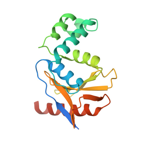
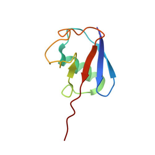
4BOZ

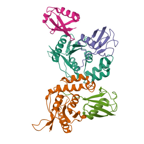
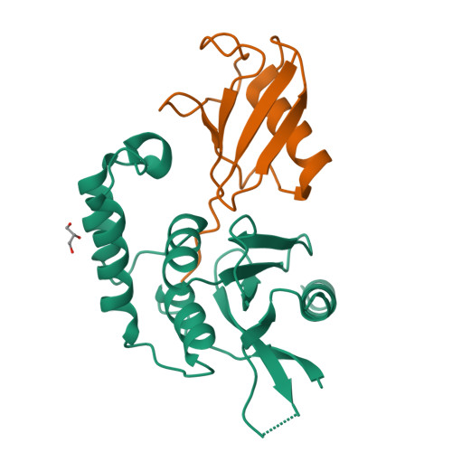
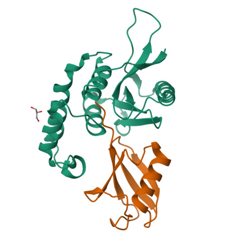
Structure of OTUD2 OTU domain in complex with K11-linked di ubiquitin


Experimental Data Snapshot

  • Method: X-RAY DIFFRACTION
  • Resolution: 3.03 Å
  • R-Value Free: 0.254 
  • R-Value Work: 0.193 
  • R-Value Observed: 0.196 

Starting Model: experimental
View more details

wwPDB Validation   3D Report Full Report


This is version 1.1 of the entry. See complete history


Literature

Otu Deubiquitinases Reveal Mechanisms of Linkage Specificity and Enable Ubiquitin Chain Restriction Analysis.

Mevissen, T.E.T.Hospenthal, M.K.Geurink, P.P.Elliott, P.R.Akutsu, M.Arnaudo, N.Ekkebus, R.Kulathu, Y.Wauer, T.El Oualid, F.Freund, S.M.V.Ovaa, H.Komander, D.

(2013) Cell 154: 169

  • DOI: https://doi.org/10.1016/j.cell.2013.05.046
  • Primary Citation of Related Structures:  
    4BOP, 4BOQ, 4BOS, 4BOU, 4BOZ

  • PubMed Abstract: 

    Sixteen ovarian tumor (OTU) family deubiquitinases (DUBs) exist in humans, and most members regulate cell-signaling cascades. Several OTU DUBs were reported to be ubiquitin (Ub) chain linkage specific, but comprehensive analyses are missing, and the underlying mechanisms of linkage specificity are unclear. Using Ub chains of all eight linkage types, we reveal that most human OTU enzymes are linkage specific, preferring one, two, or a defined subset of linkage types, including unstudied atypical Ub chains. Biochemical analysis and five crystal structures of OTU DUBs with or without Ub substrates reveal four mechanisms of linkage specificity. Additional Ub-binding domains, the ubiquitinated sequence in the substrate, and defined S1' and S2 Ub-binding sites on the OTU domain enable OTU DUBs to distinguish linkage types. We introduce Ub chain restriction analysis, in which OTU DUBs are used as restriction enzymes to reveal linkage type and the relative abundance of Ub chains on substrates.


  • Organizational Affiliation

    Medical Research Council Laboratory of Molecular Biology, Francis Crick Avenue, Cambridge Biomedical Campus, Cambridge CB2 0QH, UK.


Macromolecules
Find similar proteins by:  (by identity cutoff)  |  3D Structure
Entity ID: 1
MoleculeChains Sequence LengthOrganismDetailsImage
UBIQUITIN THIOESTERASE OTU1
A, D
183Homo sapiensMutation(s): 1 
EC: 3.4.19.12
UniProt & NIH Common Fund Data Resources
Find proteins for Q5VVQ6 (Homo sapiens)
Explore Q5VVQ6 
Go to UniProtKB:  Q5VVQ6
PHAROS:  Q5VVQ6
GTEx:  ENSG00000180667 
Entity Groups  
Sequence Clusters30% Identity50% Identity70% Identity90% Identity95% Identity100% Identity
UniProt GroupQ5VVQ6
Sequence Annotations
Expand
  • Reference Sequence
Find similar proteins by:  (by identity cutoff)  |  3D Structure
Entity ID: 2
MoleculeChains Sequence LengthOrganismDetailsImage
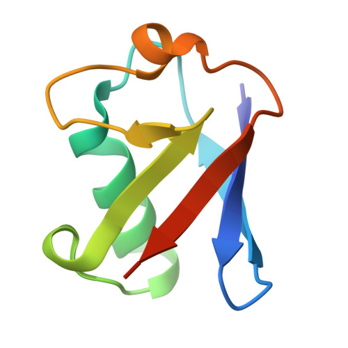
UBIQUITIN-C
B, C, E
76Homo sapiensMutation(s): 0 
UniProt & NIH Common Fund Data Resources
Find proteins for P0CG48 (Homo sapiens)
Explore P0CG48 
Go to UniProtKB:  P0CG48
PHAROS:  P0CG48
GTEx:  ENSG00000150991 
Entity Groups  
Sequence Clusters30% Identity50% Identity70% Identity90% Identity95% Identity100% Identity
UniProt GroupP0CG48
Sequence Annotations
Expand
  • Reference Sequence
Experimental Data & Validation

Experimental Data

  • Method: X-RAY DIFFRACTION
  • Resolution: 3.03 Å
  • R-Value Free: 0.254 
  • R-Value Work: 0.193 
  • R-Value Observed: 0.196 
  • Space Group: C 1 2 1
Unit Cell:
Length ( Å )Angle ( ˚ )
a = 174.79α = 90
b = 44.22β = 91.41
c = 84.95γ = 90
Software Package:
Software NamePurpose
PHENIXrefinement
MOSFLMdata reduction
SCALEPACKdata scaling
PHASERphasing

Structure Validation

View Full Validation Report


Entry History 

Revision History  (Full details and data files)

  • Version 1.0: 2013-07-17
    Type: Initial release
  • Version 1.1: 2023-12-20
    Changes: Data collection, Database references, Derived calculations, Other, Refinement description