4BMH | pdb_00004bmh

Crystal structure of SsHAT


Experimental Data Snapshot

  • Method: X-RAY DIFFRACTION
  • Resolution: 1.50 Å
  • R-Value Free: 
    0.175 (Depositor), 0.174 (DCC) 
  • R-Value Work: 
    0.146 (Depositor), 0.145 (DCC) 
  • R-Value Observed: 
    0.148 (Depositor) 

wwPDB Validation 3D Report Full Report

Validation slider image for 4BMH

This is version 1.2 of the entry. See complete history

Literature

Three-Dimensional Structure of a Streptomyces Sviceus Gnat Acetyltransferase with Similarity to the C-Terminal Domain of the Human Gh84 O-Glcnacase

He, Y.Roth, C.Turkenburg, J.P.Davies, G.J.

(2014) Acta Crystallogr D Biol Crystallogr 70: 186

  • DOI: https://doi.org/10.1107/S1399004713029155
  • Primary Citation Related Structures: 
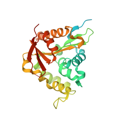
    4BMH

  • PubMed Abstract: 

    The mammalian O-GlcNAc hydrolysing enzyme O-GlcNAcase (OGA) is a multi-domain protein with glycoside hydrolase activity in the N-terminus and with a C-terminal domain that has low sequence similarity to known acetyltransferases, prompting speculation, albeit controversial, that the C-terminal domain may function as a histone acetyltransferase (HAT). There are currently scarce data available regarding the structure and function of this C-terminal region. Here, a bacterial homologue of the human OGA C-terminal domain, an acetyltransferase protein (accession No. ZP_05014886) from Streptomyces sviceus (SsAT), was cloned and its crystal structure was solved to high resolution. The structure reveals a conserved protein core that has considerable structural homology to the acetyl-CoA (AcCoA) binding site of GCN5-related acetyltransferases (GNATs). Calorimetric data further confirm that SsAT is indeed able to bind AcCoA in solution with micromolar affinity. Detailed structural analysis provided insight into the binding of AcCoA. An acceptor-binding cavity was identified, indicating that the physiological substrate of SsAT may be a small molecule. Consistent with recently published work, the SsAT structure further questions a HAT function for the human OGA domain.


  • Organizational Affiliation
    • College of Chemistry and Materials Science, Northwest University, Xi'an 710069, People's Republic of China.

Macromolecule Content 

  • Total Structure Weight: 28.71 kDa 
  • Atom Count: 1,881 
  • Modeled Residue Count: 202 
  • Deposited Residue Count: 262 
  • Unique protein chains: 1

Macromolecules

Find similar proteins by:|  3D Structure
Entity ID: 1
MoleculeChains  Sequence LengthOrganismDetailsImage
ACETYLTRANSFERASE262Streptomyces sviceus ATCC 29083Mutation(s): 0 
UniProt
Find proteins for B5HKU0 (Streptomyces sviceus (strain ATCC 29083 / DSM 924 / JCM 4929 / NBRC 13980 / NCIMB 11184 / NRRL 5439 / UC 5370))
Explore B5HKU0 
Go to UniProtKB:  B5HKU0
Entity Groups
Sequence Clusters30% Identity50% Identity70% Identity90% Identity95% Identity100% Identity
UniProt GroupB5HKU0
Sequence Annotations
Expand
Reference Sequence

Small Molecules

Ligands 1 Unique
IDChains Name / Formula / InChI Key2D Diagram3D Interactions
CL

Query on CL


Download:Ideal Coordinates CCD File
B [auth A]CHLORIDE ION
Cl
VEXZGXHMUGYJMC-UHFFFAOYSA-M

Experimental Data & Validation

Experimental Data

  • Method: X-RAY DIFFRACTION
  • Resolution: 1.50 Å
  • R-Value Free:  0.175 (Depositor), 0.174 (DCC) 
  • R-Value Work:  0.146 (Depositor), 0.145 (DCC) 
  • R-Value Observed: 0.148 (Depositor) 
Space Group: P 32 2 1
Unit Cell:
Length ( Å )Angle ( ˚ )
a = 75.885α = 90
b = 75.885β = 90
c = 76.517γ = 120
Software Package:
Software NamePurpose
REFMACrefinement
MOSFLMdata reduction
SCALAdata scaling
SHELXphasing

Structure Validation

View Full Validation Report


Entry History 

Deposition Data

Revision History  (Full details and data files)

  • Version 1.0: 2014-01-15
    Type: Initial release
  • Version 1.1: 2014-01-22
    Changes: Database references
  • Version 1.2: 2024-05-08
    Changes: Data collection, Database references, Derived calculations, Other