4BJ8 | pdb_00004bj8

Zebavidin


Experimental Data Snapshot

  • Method: X-RAY DIFFRACTION
  • Resolution: 2.40 Å
  • R-Value Free: 
    0.266 (Depositor), 0.264 (DCC) 
  • R-Value Work: 
    0.192 (Depositor), 0.198 (DCC) 
  • R-Value Observed: 
    0.195 (Depositor) 

Starting Model: experimental
View more details

wwPDB Validation 3D Report Full Report

Validation slider image for 4BJ8

Ligand Structure Quality Assessment 


This is version 1.3 of the entry. See complete history

Literature

Zebavidin

Niederhauser, B.Zmurko, J.Parthiban, M.Ojanen, M.Kukkurainen, S.Maatta, J.A.E.Leppiniemi, J.Janis, J.Parikka, M.Turpeinen, H.Pesu, M.Johnson, M.S.Airenne, T.T.Kulomaa, M.S.Hytonen, V.P.

(2013) PLoS One 8: 77207

  • DOI: https://doi.org/10.1371/journal.pone.0077207
  • Primary Citation Related Structures: 
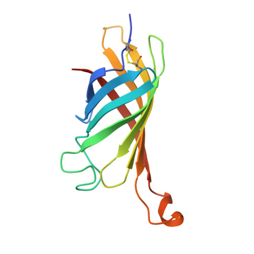
    4BJ8

  • PubMed Abstract: 

    The avidin protein family members are well known for their high affinity towards D-biotin and high structural stability. These properties make avidins valuable tools for a wide range of biotechnology applications. We have identified a new member of the avidin family in the zebrafish (Danio rerio) genome, hereafter called zebavidin. The protein is highly expressed in the gonads of both male and female zebrafish and in the gills of male fish, but our data suggest that zebavidin is not crucial for the developing embryo. Biophysical and structural characterisation of zebavidin revealed distinct properties not found in any previously characterised avidins. Gel filtration chromatography and native mass spectrometry suggest that the protein forms dimers in the absence of biotin at low ionic strength, but assembles into tetramers upon binding biotin. Ligand binding was analysed using radioactive and fluorescently labelled biotin and isothermal titration calorimetry. Moreover, the crystal structure of zebavidin in complex with biotin was solved at 2.4 Å resolution and unveiled unique ligand binding and subunit interface architectures; the atomic-level details support our physicochemical observations.


  • Organizational Affiliation
    • Institute of Biomedical Technology, University of Tampere, BioMediTech, Tampere, Finland ; Fimlab Laboratories, Pirkanmaa Hospital District, Tampere, Finland.

Macromolecule Content 

  • Total Structure Weight: 221.69 kDa 
  • Atom Count: 15,389 
  • Modeled Residue Count: 1,908 
  • Deposited Residue Count: 2,016 
  • Unique protein chains: 1

Macromolecules

Find similar proteins by:|  3D Structure
Entity ID: 1
MoleculeChains  Sequence LengthOrganismDetailsImage
ZEBAVIDIN
A, B, C, D, E
A, B, C, D, E, F, G, H, I, J, K, L, M, N, O, P
126Danio rerioMutation(s): 0 
UniProt
Find proteins for E7F650 (Danio rerio)
Explore E7F650 
Go to UniProtKB:  E7F650
Entity Groups
Sequence Clusters30% Identity50% Identity70% Identity90% Identity95% Identity100% Identity
UniProt GroupE7F650
Sequence Annotations
Expand
Reference Sequence

Small Molecules

Ligands 2 Unique
IDChains Name / Formula / InChI Key2D Diagram3D Interactions
BTN

Query on BTN


Download:Ideal Coordinates CCD File
AA [auth K]
BA [auth L]
DA [auth M]
EA [auth N]
FA [auth O]
AA [auth K],
BA [auth L],
DA [auth M],
EA [auth N],
FA [auth O],
GA [auth P],
Q [auth A],
R [auth B],
S [auth C],
T [auth D],
U [auth E],
V [auth F],
W [auth G],
X [auth H],
Y [auth I],
Z [auth J]
BIOTIN
C10 H16 N2 O3 S
YBJHBAHKTGYVGT-ZKWXMUAHSA-N
GOL

Query on GOL


Download:Ideal Coordinates CCD File
CA [auth L]GLYCEROL
C3 H8 O3
PEDCQBHIVMGVHV-UHFFFAOYSA-N

Experimental Data & Validation

Experimental Data

  • Method: X-RAY DIFFRACTION
  • Resolution: 2.40 Å
  • R-Value Free:  0.266 (Depositor), 0.264 (DCC) 
  • R-Value Work:  0.192 (Depositor), 0.198 (DCC) 
  • R-Value Observed: 0.195 (Depositor) 
Space Group: P 21 21 2
Unit Cell:
Length ( Å )Angle ( ˚ )
a = 182.24α = 90
b = 196.84β = 90
c = 52.59γ = 90
Software Package:
Software NamePurpose
REFMACrefinement
XDSdata reduction
XSCALEdata scaling
PHASERphasing

Structure Validation

View Full Validation Report


Ligand Structure Quality Assessment 


Entry History 

Deposition Data

Revision History  (Full details and data files)

  • Version 1.0: 2013-11-20
    Type: Initial release
  • Version 1.1: 2019-05-08
    Changes: Data collection, Experimental preparation, Other
  • Version 1.2: 2023-12-20
    Changes: Data collection, Database references, Derived calculations, Other, Refinement description
  • Version 1.3: 2024-10-23
    Changes: Structure summary