4BJ4 | pdb_00004bj4

Structure of Pseudomonas aeruginosa amidase Ampdh2


Experimental Data Snapshot

  • Method: X-RAY DIFFRACTION
  • Resolution: 1.72 Å
  • R-Value Free: 
    0.201 (Depositor), 0.201 (DCC) 
  • R-Value Work: 
    0.172 (Depositor), 0.173 (DCC) 
  • R-Value Observed: 
    0.173 (Depositor) 

Starting Model: experimental
View more details

wwPDB Validation 3D Report Full Report

Validation slider image for 4BJ4

This is version 1.2 of the entry. See complete history

Literature

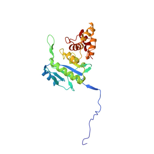
Reaction Products and the X-Ray Structure of Ampdh2, a Virulence Determinant of Pseudomonas Aeruginosa.

Martinez-Caballero, S.Lee, M.Artola-Recolons, C.Carrasco-Lopez, C.Hesek, D.Spink, E.E.Lastochkin, E.Zhang, W.Hellman, L.M.Boggess, B.Mobashery, S.Hermoso, J.A.

(2013) J Am Chem Soc 135: 10318

  • DOI: https://doi.org/10.1021/ja405464b
  • Primary Citation Related Structures: 
    4BJ4, 4BOL, 4BPA

  • PubMed Abstract: 

    The zinc protease AmpDh2 is a virulence determinant of Pseudomonas aeruginosa, a problematic human pathogen. The mechanism of how the protease manifests virulence is not known, but it is known that it turns over the bacterial cell wall. The reaction of AmpDh2 with the cell wall was investigated, and nine distinct turnover products were characterized by LC/MS/MS. The enzyme turns over both the cross-linked and noncross-linked cell wall. Three high-resolution X-ray structures, the apo enzyme and two complexes with turnover products, were solved. The X-ray structures show how the dimeric protein interacts with the inner leaflet of the bacterial outer membrane and that the two monomers provide a more expansive surface for recognition of the cell wall. This binding surface can accommodate the 3D solution structure of the cross-linked cell wall.


  • Organizational Affiliation
    • Department of Crystallography and Structural Biology, Inst. Química-Física "Rocasolano", CSIC, Serrano 119, 28006 Madrid, Spain.

Macromolecule Content 

  • Total Structure Weight: 55.18 kDa 
  • Atom Count: 4,799 
  • Modeled Residue Count: 492 
  • Deposited Residue Count: 492 
  • Unique protein chains: 1

Macromolecules

Find similar proteins by:|  3D Structure
Entity ID: 1
MoleculeChains  Sequence LengthOrganismDetailsImage
AMPDH2
A, B
246Pseudomonas aeruginosa PAO1Mutation(s): 0 
EC: 3.5.1.28
UniProt
Find proteins for Q9HT86 (Pseudomonas aeruginosa (strain ATCC 15692 / DSM 22644 / CIP 104116 / JCM 14847 / LMG 12228 / 1C / PRS 101 / PAO1))
Explore Q9HT86 
Go to UniProtKB:  Q9HT86
Entity Groups
Sequence Clusters30% Identity50% Identity70% Identity90% Identity95% Identity100% Identity
UniProt GroupQ9HT86
Sequence Annotations
Expand
Reference Sequence

Small Molecules

Ligands 1 Unique
IDChains Name / Formula / InChI Key2D Diagram3D Interactions
FLC

Query on FLC


Download:Ideal Coordinates CCD File
C [auth A]CITRATE ANION
C6 H5 O7
KRKNYBCHXYNGOX-UHFFFAOYSA-K

Experimental Data & Validation

Experimental Data

  • Method: X-RAY DIFFRACTION
  • Resolution: 1.72 Å
  • R-Value Free:  0.201 (Depositor), 0.201 (DCC) 
  • R-Value Work:  0.172 (Depositor), 0.173 (DCC) 
  • R-Value Observed: 0.173 (Depositor) 
Space Group: P 21 21 21
Unit Cell:
Length ( Å )Angle ( ˚ )
a = 48.759α = 90
b = 100.421β = 90
c = 104.26γ = 90
Software Package:
Software NamePurpose
PHENIXrefinement
XDSdata reduction
SCALAdata scaling

Structure Validation

View Full Validation Report


Entry History 

Deposition Data

Revision History  (Full details and data files)

  • Version 1.0: 2013-07-24
    Type: Initial release
  • Version 1.1: 2013-07-31
    Changes: Database references
  • Version 1.2: 2023-12-20
    Changes: Data collection, Database references, Derived calculations, Other, Refinement description