4BEM

Crystal structure of the F-type ATP synthase c-ring from Acetobacterium woodii.


Experimental Data Snapshot

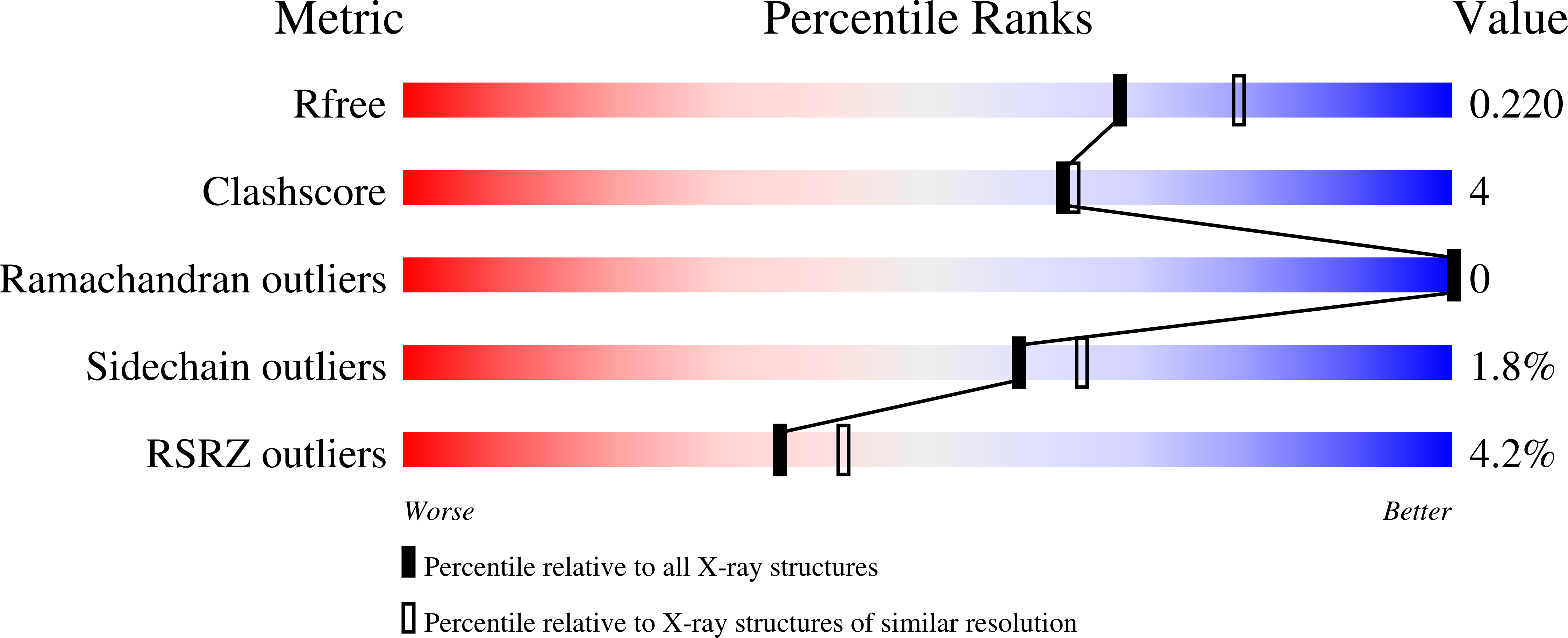
  • Method: X-RAY DIFFRACTION
  • Resolution: 2.10 Å
  • R-Value Free: 0.220 
  • R-Value Work: 0.182 
  • R-Value Observed: 0.184 

Starting Model: experimental
View more details

wwPDB Validation   3D Report Full Report


Ligand Structure Quality Assessment 


This is version 1.5 of the entry. See complete history


Literature

High-Resolution Structure and Mechanism of an F/V-Hybrid Rotor Ring in a Na+-Coupled ATP Synthase

Matthies, D.Zhou, W.Klyszejko, A.L.Anselmi, C.Yildiz, O.Brandt, K.Muller, V.Faraldo-Gomez, J.D.Meier, T.

(2014) Nat Commun 5: 5286

  • DOI: https://doi.org/10.1038/ncomms6286
  • Primary Citation of Related Structures:  
    4BEM

  • PubMed Abstract: 

    All rotary ATPases catalyse the interconversion of ATP and ADP-Pi through a mechanism that is coupled to the transmembrane flow of H(+) or Na(+). Physiologically, however, F/A-type enzymes specialize in ATP synthesis driven by downhill ion diffusion, while eukaryotic V-type ATPases function as ion pumps. To begin to rationalize the molecular basis for this functional differentiation, we solved the crystal structure of the Na(+)-driven membrane rotor of the Acetobacterium woodii ATP synthase, at 2.1 Å resolution. Unlike known structures, this rotor ring is a 9:1 heteromer of F- and V-type c-subunits and therefore features a hybrid configuration of ion-binding sites along its circumference. Molecular and kinetic simulations are used to dissect the mechanisms of Na(+) recognition and rotation of this c-ring, and to explain the functional implications of the V-type c-subunit. These structural and mechanistic insights indicate an evolutionary path between synthases and pumps involving adaptations in the rotor ring.


  • Organizational Affiliation

    Department of Structural Biology, Max Planck Institute of Biophysics, Max-von-Laue-Str. 3, 60438 Frankfurt am Main, Germany.


Macromolecules
Find similar proteins by:  (by identity cutoff)  |  3D Structure
Entity ID: 1
MoleculeChains Sequence LengthOrganismDetailsImage
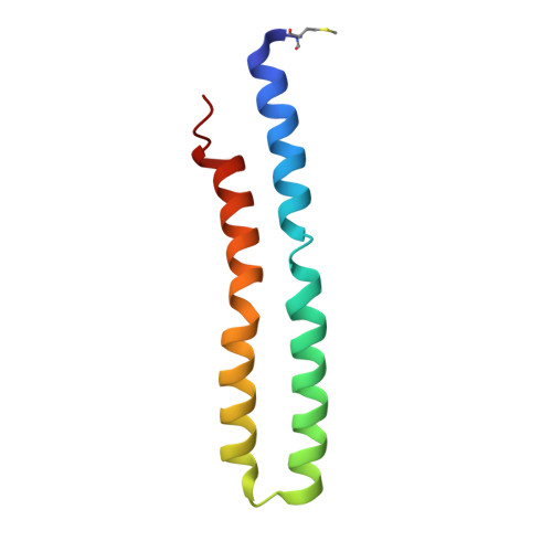
F1FO ATPASE C2 SUBUNIT
A, B, C, D, E
A, B, C, D, E, F, G, H, I
82Acetobacterium woodii DSM 1030Mutation(s): 0 
Membrane Entity: Yes 
UniProt
Find proteins for H6LFT2 (Acetobacterium woodii (strain ATCC 29683 / DSM 1030 / JCM 2381 / KCTC 1655 / WB1))
Explore H6LFT2 
Go to UniProtKB:  H6LFT2
Entity Groups  
Sequence Clusters30% Identity50% Identity70% Identity90% Identity95% Identity100% Identity
UniProt GroupH6LFT2
Sequence Annotations
Expand
  • Reference Sequence
Find similar proteins by:  (by identity cutoff)  |  3D Structure
Entity ID: 2
MoleculeChains Sequence LengthOrganismDetailsImage
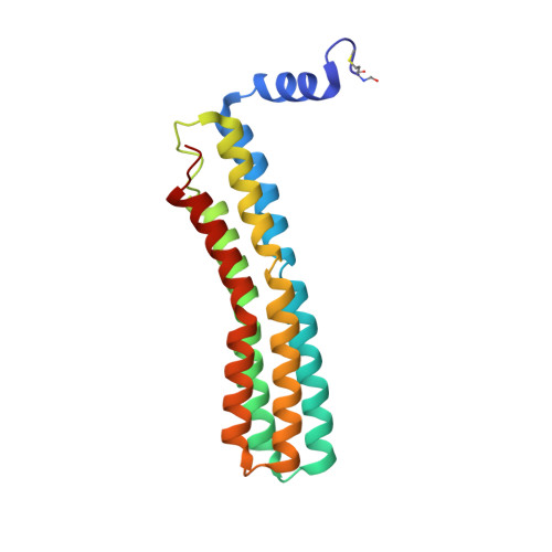
F1FO ATPASE C1 SUBUNIT182Acetobacterium woodii DSM 1030Mutation(s): 0 
Membrane Entity: Yes 
UniProt
Find proteins for H6LFT0 (Acetobacterium woodii (strain ATCC 29683 / DSM 1030 / JCM 2381 / KCTC 1655 / WB1))
Explore H6LFT0 
Go to UniProtKB:  H6LFT0
Entity Groups  
Sequence Clusters30% Identity50% Identity70% Identity90% Identity95% Identity100% Identity
UniProt GroupH6LFT0
Sequence Annotations
Expand
  • Reference Sequence
Small Molecules
Ligands 6 Unique
IDChains Name / Formula / InChI Key2D Diagram3D Interactions
HTG
Query on HTG

Download Ideal Coordinates CCD File 
AA [auth G]
BA [auth G]
CA [auth G]
EA [auth H]
FA [auth H]
AA [auth G],
BA [auth G],
CA [auth G],
EA [auth H],
FA [auth H],
JA [auth I],
KA [auth I],
LA [auth I],
M [auth B],
MA [auth I],
OA [auth J],
PA [auth J],
QA [auth J],
RA [auth J],
SA [auth J],
T [auth E],
TA [auth J],
U [auth E],
W [auth F]
heptyl 1-thio-beta-D-glucopyranoside
C13 H26 O5 S
HPEGNLMTTNTJSP-LBELIVKGSA-N
P6G
Query on P6G

Download Ideal Coordinates CCD File 
S [auth E]HEXAETHYLENE GLYCOL
C12 H26 O7
IIRDTKBZINWQAW-UHFFFAOYSA-N
TAM
Query on TAM

Download Ideal Coordinates CCD File 
P [auth C]TRIS(HYDROXYETHYL)AMINOMETHANE
C7 H17 N O3
GKODZWOPPOTFGA-UHFFFAOYSA-N
ACT
Query on ACT

Download Ideal Coordinates CCD File 
HA [auth I],
IA [auth I],
X [auth F],
Z [auth G]
ACETATE ION
C2 H3 O2
QTBSBXVTEAMEQO-UHFFFAOYSA-M
MN
Query on MN

Download Ideal Coordinates CCD File 
O [auth C]MANGANESE (II) ION
Mn
WAEMQWOKJMHJLA-UHFFFAOYSA-N
NA
Query on NA

Download Ideal Coordinates CCD File 
DA [auth H]
GA [auth I]
K [auth A]
L [auth B]
N [auth C]
DA [auth H],
GA [auth I],
K [auth A],
L [auth B],
N [auth C],
NA [auth J],
Q [auth D],
R [auth E],
V [auth F],
Y [auth G]
SODIUM ION
Na
FKNQFGJONOIPTF-UHFFFAOYSA-N
Modified Residues  1 Unique
IDChains TypeFormula2D DiagramParent
FME
Query on FME
A, B, C, D, E
A, B, C, D, E, F, G, H, I
L-PEPTIDE LINKINGC6 H11 N O3 SMET
Experimental Data & Validation

Experimental Data

  • Method: X-RAY DIFFRACTION
  • Resolution: 2.10 Å
  • R-Value Free: 0.220 
  • R-Value Work: 0.182 
  • R-Value Observed: 0.184 
  • Space Group: P 43 21 2
Unit Cell:
Length ( Å )Angle ( ˚ )
a = 121.17α = 90
b = 121.17β = 90
c = 150.57γ = 90
Software Package:
Software NamePurpose
PHENIXrefinement
XDSdata reduction
XSCALEdata scaling
PHASERphasing

Structure Validation

View Full Validation Report


Ligand Structure Quality Assessment 


Entry History 

Deposition Data

Revision History  (Full details and data files)

  • Version 1.0: 2014-03-26
    Type: Initial release
  • Version 1.1: 2014-10-01
    Changes: Database references
  • Version 1.2: 2014-11-19
    Changes: Database references
  • Version 1.3: 2020-07-29
    Type: Remediation
    Reason: Carbohydrate remediation
    Changes: Derived calculations, Other, Structure summary
  • Version 1.4: 2023-12-20
    Changes: Data collection, Database references, Refinement description, Structure summary
  • Version 1.5: 2024-10-23
    Changes: Structure summary