4BD6 | pdb_00004bd6

Bax domain swapped dimer in complex with BaxBH3


Experimental Data Snapshot

  • Method: X-RAY DIFFRACTION
  • Resolution: 2.49 Å
  • R-Value Free: 
    0.285 (Depositor), 0.293 (DCC) 
  • R-Value Work: 
    0.246 (Depositor), 0.260 (DCC) 
  • R-Value Observed: 
    0.248 (Depositor) 

Starting Model: other
View more details

wwPDB Validation 3D Report Full Report

Validation slider image for 4BD6

This is version 1.1 of the entry. See complete history

Literature

Bax Crystal Structures Reveal How Bh3 Domains Activate Bax and Nucleate its Oligomerization to Induce Apoptosis.

Czabotar, P.E.Westphal, D.Dewson, G.Ma, S.Hockings, C.Fairlie, W.D.Lee, E.F.Yao, S.Robin, A.Y.Smith, B.J.Huang, D.C.Kluck, R.M.Adams, J.M.Colman, P.M.

(2013) Cell 152: 519

  • DOI: https://doi.org/10.1016/j.cell.2012.12.031
  • Primary Citation Related Structures: 
    4BD2, 4BD6, 4BD7, 4BD8, 4BDU

  • PubMed Abstract: 

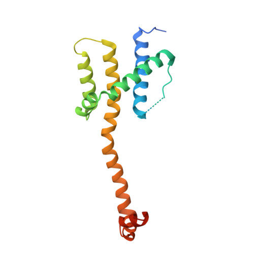
    In stressed cells, apoptosis ensues when Bcl-2 family members Bax or Bak oligomerize and permeabilize the mitochondrial outer membrane. Certain BH3-only relatives can directly activate them to mediate this pivotal, poorly understood step. To clarify the conformational changes that induce Bax oligomerization, we determined crystal structures of BaxΔC21 treated with detergents and BH3 peptides. The peptides bound the Bax canonical surface groove but, unlike their complexes with prosurvival relatives, dissociated Bax into two domains. The structures define the sequence signature of activator BH3 domains and reveal how they can activate Bax via its groove by favoring release of its BH3 domain. Furthermore, Bax helices α2-α5 alone adopted a symmetric homodimer structure, supporting the proposal that two Bax molecules insert their BH3 domain into each other's surface groove to nucleate oligomerization. A planar lipophilic surface on this homodimer may engage the membrane. Our results thus define critical Bax transitions toward apoptosis.


  • Organizational Affiliation
    • The Walter and Eliza Hall Institute of Medical Research, Melbourne, Victoria 3052, Australia. czabotar@wehi.edu.au

Macromolecule Content 

  • Total Structure Weight: 23.02 kDa 
  • Atom Count: 1,314 
  • Modeled Residue Count: 167 
  • Deposited Residue Count: 208 
  • Unique protein chains: 2

Macromolecules

Find similar proteins by:|  3D Structure
Entity ID: 1
MoleculeChains  Sequence LengthOrganismDetailsImage
APOPTOSIS REGULATOR BAX174Homo sapiensMutation(s): 2 
UniProt & NIH Common Fund Data Resources
Find proteins for Q07812 (Homo sapiens)
Explore Q07812 
Go to UniProtKB:  Q07812
PHAROS:  Q07812
GTEx:  ENSG00000087088 
Entity Groups
Sequence Clusters30% Identity50% Identity70% Identity90% Identity95% Identity100% Identity
UniProt GroupQ07812
Sequence Annotations
Expand
Reference Sequence
Find similar proteins by:|  3D Structure
Entity ID: 2
MoleculeChains  Sequence LengthOrganismDetailsImage
APOPTOSIS REGULATOR BAXB [auth C]34Homo sapiensMutation(s): 0 
UniProt & NIH Common Fund Data Resources
Find proteins for Q07812 (Homo sapiens)
Explore Q07812 
Go to UniProtKB:  Q07812
PHAROS:  Q07812
GTEx:  ENSG00000087088 
Entity Groups
Sequence Clusters30% Identity50% Identity70% Identity90% Identity95% Identity100% Identity
UniProt GroupQ07812
Sequence Annotations
Expand
Reference Sequence

Experimental Data & Validation

Experimental Data

  • Method: X-RAY DIFFRACTION
  • Resolution: 2.49 Å
  • R-Value Free:  0.285 (Depositor), 0.293 (DCC) 
  • R-Value Work:  0.246 (Depositor), 0.260 (DCC) 
  • R-Value Observed: 0.248 (Depositor) 
Space Group: P 43 21 2
Unit Cell:
Length ( Å )Angle ( ˚ )
a = 103.715α = 90
b = 103.715β = 90
c = 40.555γ = 90
Software Package:
Software NamePurpose
PHENIXrefinement
XDSdata reduction
XDSdata scaling
PHASERphasing

Structure Validation

View Full Validation Report


Entry History 

Deposition Data

Revision History  (Full details and data files)

  • Version 1.0: 2013-02-13
    Type: Initial release
  • Version 1.1: 2024-05-01
    Changes: Data collection, Database references, Other, Refinement description