4BBJ | pdb_00004bbj

Copper-transporting PIB-ATPase in complex with beryllium fluoride representing the E2P state


Experimental Data Snapshot

  • Method: X-RAY DIFFRACTION
  • Resolution: 2.75 Å
  • R-Value Free: 
    0.247 (Depositor) 
  • R-Value Work: 
    0.199 (Depositor) 
  • R-Value Observed: 
    0.202 (Depositor) 

Starting Model: experimental
View more details

wwPDB Validation 3D Report Full Report

Validation slider image for 4BBJ

Ligand Structure Quality Assessment 


This is version 1.3 of the entry. See complete history

Literature

Copper-Transporting P-Type Atpases Use a Unique Ion-Release Pathway

Andersson, M.Mattle, D.Sitsel, O.Klymchuk, T.Nielsen, A.Moller, L.B.White, S.H.Nissen, P.Gourdon, P.

(2014) Nat Struct Mol Biol 21: 43

  • DOI: https://doi.org/10.1038/nsmb.2721
  • Primary Citation Related Structures: 
    4BBJ

  • PubMed Abstract: 

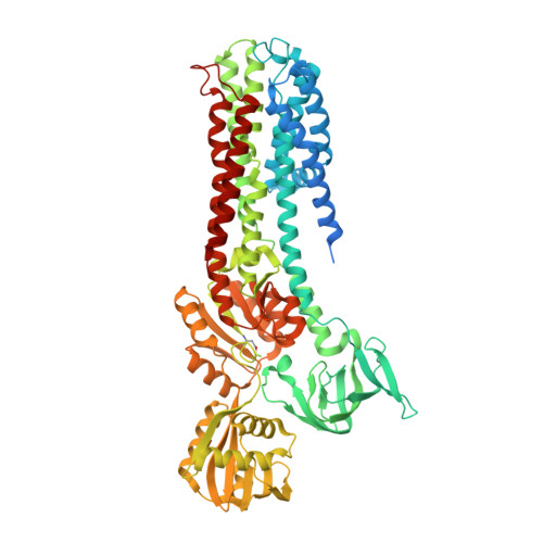
    Heavy metals in cells are typically regulated by PIB-type ATPases. The first structure of the class, a Cu(+)-ATPase from Legionella pneumophila (LpCopA), outlined a copper transport pathway across the membrane, which was inferred to be occluded. Here we show by molecular dynamics simulations that extracellular water solvated the transmembrane (TM) domain, results indicative of a Cu(+)-release pathway. Furthermore, a new LpCopA crystal structure determined at 2.8-Å resolution, trapped in the preceding E2P state, delineated the same passage, and site-directed-mutagenesis activity assays support a functional role for the conduit. The structural similarities between the TM domains of the two conformations suggest that Cu(+)-ATPases couple dephosphorylation and ion extrusion differently than do the well-characterized PII-type ATPases. The ion pathway explains why certain Menkes' and Wilson's disease mutations impair protein function and points to a site for inhibitors targeting pathogens.


  • Organizational Affiliation
    • 1] Department of Physiology and Biophysics, University of California at Irvine, Irvine, California, USA. [2] [3].

Macromolecule Content 

  • Total Structure Weight: 79.81 kDa 
  • Atom Count: 5,096 
  • Modeled Residue Count: 664 
  • Deposited Residue Count: 736 
  • Unique protein chains: 1

Macromolecules

Find similar proteins by:|  3D Structure
Entity ID: 1
MoleculeChains  Sequence LengthOrganismDetailsImage
COPPER EFFLUX ATPASE736Legionella pneumophila subsp. pneumophilaMutation(s): 0 
EC: 3.6.3 (PDB Primary Data), 7.2.2.8 (UniProt)
Membrane Entity: Yes 
UniProt
Find proteins for Q5ZWR1 (Legionella pneumophila subsp. pneumophila (strain Philadelphia 1 / ATCC 33152 / DSM 7513))
Explore Q5ZWR1 
Go to UniProtKB:  Q5ZWR1
Entity Groups
Sequence Clusters30% Identity50% Identity70% Identity90% Identity95% Identity100% Identity
UniProt GroupQ5ZWR1
Sequence Annotations
Expand
Reference Sequence

Small Molecules

Modified Residues  1 Unique
IDChains TypeFormula2D DiagramParent
BFD
Query on BFD
A
L-PEPTIDE LINKINGC4 H6 Be F3 N O4ASP

Experimental Data & Validation

Experimental Data

  • Method: X-RAY DIFFRACTION
  • Resolution: 2.75 Å
  • R-Value Free:  0.247 (Depositor) 
  • R-Value Work:  0.199 (Depositor) 
  • R-Value Observed: 0.202 (Depositor) 
Space Group: C 1 2 1
Unit Cell:
Length ( Å )Angle ( ˚ )
a = 242.01α = 90
b = 71.37β = 100.01
c = 72.43γ = 90
Software Package:
Software NamePurpose
PHENIXrefinement
XDSdata reduction
XSCALEdata scaling
PHASERphasing

Structure Validation

View Full Validation Report


Ligand Structure Quality Assessment 


Entry History 

Deposition Data

Revision History  (Full details and data files)

  • Version 1.0: 2013-12-11
    Type: Initial release
  • Version 1.1: 2015-04-15
    Changes: Structure summary
  • Version 1.2: 2023-12-20
    Changes: Data collection, Database references, Derived calculations, Other, Refinement description
  • Version 1.3: 2024-11-06
    Changes: Structure summary