4B8S | pdb_00004b8s

Crystal Structure of Thermococcus litoralis ADP-dependent Glucokinase (GK)


Experimental Data Snapshot

  • Method: X-RAY DIFFRACTION
  • Resolution: 2.58 Å
  • R-Value Free: 
    0.221 (Depositor), 0.221 (DCC) 
  • R-Value Work: 
    0.169 (Depositor), 0.170 (DCC) 
  • R-Value Observed: 
    0.172 (Depositor) 

Starting Model: experimental
View more details

wwPDB Validation 3D Report Full Report

Validation slider image for 4B8S

Ligand Structure Quality Assessment 


This is version 1.2 of the entry. See complete history

Literature

Crystal Structure, Saxs and Kinetic Mechanism of Hyperthermophilic Adp-Dependent Glucokinase from Thermococcus Litoralis Reveal a Conserved Mechanism for Catalysis.

Rivas-Pardo, J.A.Herrera-Morande, A.Castro-Fernandez, V.Fernandez, F.J.Vega, M.C.Guixe, V.

(2013) PLoS One 8: 66687

  • DOI: https://doi.org/10.1371/journal.pone.0066687
  • Primary Citation Related Structures: 
    4B8R, 4B8S

  • PubMed Abstract: 

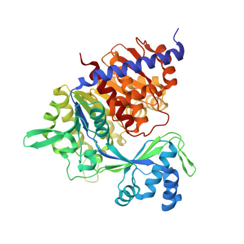
    ADP-dependent glucokinases represent a unique family of kinases that belong to the ribokinase superfamily, being present mainly in hyperthermophilic archaea. For these enzymes there is no agreement about the magnitude of the structural transitions associated with ligand binding and whether they are meaningful to the function of the enzyme. We used the ADP-dependent glucokinase from Thermococcus litoralis as a model to investigate the conformational changes observed in X-ray crystallographic structures upon substrate binding and to compare them with those determined in solution in order to understand their interplay with the glucokinase function. Initial velocity studies indicate that catalysis follows a sequential ordered mechanism that correlates with the structural transitions experienced by the enzyme in solution and in the crystal state. The combined data allowed us to resolve the open-closed conformational transition that accounts for the complete reaction cycle and to identify the corresponding clusters of aminoacids residues responsible for it. These results provide molecular bases for a general mechanism conserved across the ADP-dependent kinase family.


  • Organizational Affiliation
    • Departamento de Biología, Facultad de Ciencias, Universidad de Chile, Santiago, Chile.

Macromolecule Content 

  • Total Structure Weight: 54.68 kDa 
  • Atom Count: 4,038 
  • Modeled Residue Count: 466 
  • Deposited Residue Count: 467 
  • Unique protein chains: 1

Macromolecules

Find similar proteins by:|  3D Structure
Entity ID: 1
MoleculeChains  Sequence LengthOrganismDetailsImage
ADP-DEPENDENT GLUCOKINASE467Thermococcus litoralisMutation(s): 0 
EC: 2.7.1.147 (PDB Primary Data), 2.7.1 (UniProt)
UniProt
Find proteins for Q7M537 (Thermococcus litoralis (strain ATCC 51850 / DSM 5473 / JCM 8560 / NS-C))
Explore Q7M537 
Go to UniProtKB:  Q7M537
Entity Groups
Sequence Clusters30% Identity50% Identity70% Identity90% Identity95% Identity100% Identity
UniProt GroupQ7M537
Sequence Annotations
Expand
Reference Sequence

Small Molecules

Ligands 3 Unique
IDChains Name / Formula / InChI Key2D Diagram3D Interactions
AMP

Query on AMP


Download:Ideal Coordinates CCD File
G [auth A]ADENOSINE MONOPHOSPHATE
C10 H14 N5 O7 P
UDMBCSSLTHHNCD-KQYNXXCUSA-N
GLC

Query on GLC


Download:Ideal Coordinates CCD File
B [auth A]alpha-D-glucopyranose
C6 H12 O6
WQZGKKKJIJFFOK-DVKNGEFBSA-N
GOL

Query on GOL


Download:Ideal Coordinates CCD File
C [auth A],
D [auth A],
E [auth A],
F [auth A],
H [auth A]
GLYCEROL
C3 H8 O3
PEDCQBHIVMGVHV-UHFFFAOYSA-N

Experimental Data & Validation

Experimental Data

  • Method: X-RAY DIFFRACTION
  • Resolution: 2.58 Å
  • R-Value Free:  0.221 (Depositor), 0.221 (DCC) 
  • R-Value Work:  0.169 (Depositor), 0.170 (DCC) 
  • R-Value Observed: 0.172 (Depositor) 
Space Group: P 32 2 1
Unit Cell:
Length ( Å )Angle ( ˚ )
a = 106.468α = 90
b = 106.468β = 90
c = 130.093γ = 120
Software Package:
Software NamePurpose
REFMACrefinement
XDSdata reduction
SCALAdata scaling
PHASERphasing

Structure Validation

View Full Validation Report


Ligand Structure Quality Assessment 


Entry History 

Deposition Data

Revision History  (Full details and data files)

  • Version 1.0: 2013-07-10
    Type: Initial release
  • Version 1.1: 2020-07-29
    Type: Remediation
    Reason: Carbohydrate remediation
    Changes: Data collection, Derived calculations, Other, Structure summary
  • Version 1.2: 2023-12-20
    Changes: Data collection, Database references, Refinement description, Structure summary