


Probing the Active Center of Catalase-Phenol Oxidase from Scytalidium Thermophilum
Yuzugullu, Y., Trinh, C.H., Pearson, A.R., Ogel, Z.B., McPherson, M.J.To be published.
Experimental Data Snapshot
Entity ID: 1 | |||||
|---|---|---|---|---|---|
| Molecule | Chains | Sequence Length | Organism | Details | Image |
| CATALASE-PHENOL OXIDASE | 719 | Mycothermus thermophilus | Mutation(s): 0 EC: 1.11.1.6 | 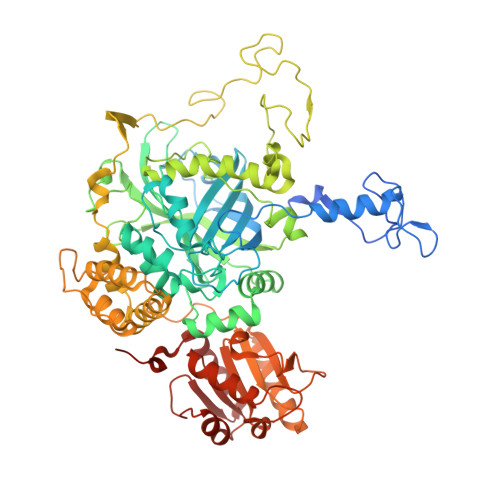 | |
UniProt | |||||
Entity Groups | |||||
| Sequence Clusters | 30% Identity50% Identity70% Identity90% Identity95% Identity100% Identity | ||||
| UniProt Group | M4GGR5 | ||||
Sequence AnnotationsExpand | |||||
Reference Sequence | |||||
| Ligands 3 Unique | |||||
|---|---|---|---|---|---|
| ID | Chains | Name / Formula / InChI Key | 2D Diagram | 3D Interactions | |
| HDD Download:Ideal Coordinates CCD File | E [auth A], J [auth B], N [auth C], R [auth D] | CIS-HEME D HYDROXYCHLORIN GAMMA-SPIROLACTONE C34 H32 Fe N4 O5 UMGOPAWIGKFTRK-QQDQPIDJSA-N |  | ||
| 3TR Download:Ideal Coordinates CCD File | F [auth A] G [auth A] K [auth B] L [auth B] O [auth C] | 3-AMINO-1,2,4-TRIAZOLE C2 H4 N4 KLSJWNVTNUYHDU-UHFFFAOYSA-N |  | ||
| CA Download:Ideal Coordinates CCD File | H [auth A], I [auth A], M [auth B], Q [auth C], U [auth D] | CALCIUM ION Ca BHPQYMZQTOCNFJ-UHFFFAOYSA-N |  | ||
| Length ( Å ) | Angle ( ˚ ) |
|---|---|
| a = 200.865 | α = 90 |
| b = 121.677 | β = 115.5 |
| c = 125.528 | γ = 90 |
| Software Name | Purpose |
|---|---|
| REFMAC | refinement |
| XDS | data reduction |
| SCALA | data scaling |


