4B52 | pdb_00004b52

Crystal structure of Gentlyase, the neutral metalloprotease of Paenibacillus polymyxa


Experimental Data Snapshot

  • Method: X-RAY DIFFRACTION
  • Resolution: 1.76 Å
  • R-Value Free: 
    0.247 (Depositor), 0.248 (DCC) 
  • R-Value Work: 
    0.195 (Depositor), 0.195 (DCC) 
  • R-Value Observed: 
    0.198 (Depositor) 

Starting Model: experimental
View more details

wwPDB Validation 3D Report Full Report

Validation slider image for 4B52

Ligand Structure Quality Assessment 


This is version 1.5 of the entry. See complete history

Literature

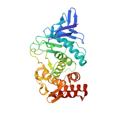
Structure of Gentlyase, the Neutral Metalloprotease of Paenibacillus Polymyxa

Ruf, A.Stihle, M.Benz, J.Schmidt, M.Sobek, H.

(2013) Acta Crystallogr D Biol Crystallogr 69: 24

  • DOI: https://doi.org/10.1107/S0907444912041169
  • Primary Citation Related Structures: 
    4B52, 4GER

  • PubMed Abstract: 

    Gentlyase is a bacterial extracellular metalloprotease that is widely applied in cell culture and for tissue dissociation and that belongs to the family of thermolysin-like proteases. The structure of thermolysin has been known since 1972 and that of Bacillus cereus neutral protease since 1992. However, the structure determination of other Bacillus neutral proteases has been hindered by their tendency to cannibalistic autolysis. High calcium conditions that allow the concentration and crystallization of the active Gentlyase metalloprotease without autoproteolysis were identified using thermal fluorescent shift assays. X-ray structures of the protease were solved in the absence and in the presence of the inhibitor phosphoramidon at 1.59 and 1.76 Å resolution, respectively. No domain movement was observed upon inhibitor binding, although such movement is thought to be a general feature of the thermolysin-like protease family. Further analysis of the structure shows that the observed calcium dependency of Gentlyase stability may arise from a partly degenerated calcium site Ca1-2 and a deletion near site Ca3.


  • Organizational Affiliation
    • pRED Pharma Research and Early Development, Small Molecule Research, Discovery Technologies, F. Hoffmann-La Roche Ltd, Basel, Switzerland. armin.ruf@roche.com

Macromolecule Content 

  • Total Structure Weight: 66.3 kDa 
  • Atom Count: 5,295 
  • Modeled Residue Count: 608 
  • Deposited Residue Count: 608 
  • Unique protein chains: 1

Macromolecules

Find similar proteins by:|  3D Structure
Entity ID: 1
MoleculeChains  Sequence LengthOrganismDetailsImage
BACILLOLYSIN
A, B
304Paenibacillus polymyxaMutation(s): 0 
EC: 3.4.24.4 (PDB Primary Data), 3.4.24 (UniProt)
UniProt
Find proteins for E3E6L0 (Paenibacillus polymyxa (strain SC2))
Explore E3E6L0 
Go to UniProtKB:  E3E6L0
Entity Groups
Sequence Clusters30% Identity50% Identity70% Identity90% Identity95% Identity100% Identity
UniProt GroupE3E6L0
Sequence Annotations
Expand
Reference Sequence

Small Molecules

Ligands 4 Unique
IDChains Name / Formula / InChI Key2D Diagram3D Interactions
RDF

Query on RDF


Download:Ideal Coordinates CCD File
G [auth A],
M [auth B]
N-ALPHA-L-RHAMNOPYRANOSYLOXY(HYDROXYPHOSPHINYL)-L-LEUCYL-L-TRYPTOPHAN
C23 H34 N3 O10 P
ZPHBZEQOLSRPAK-XLCYBJAPSA-N
ZN

Query on ZN


Download:Ideal Coordinates CCD File
C [auth A],
I [auth B]
ZINC ION
Zn
PTFCDOFLOPIGGS-UHFFFAOYSA-N
CA

Query on CA


Download:Ideal Coordinates CCD File
D [auth A]
E [auth A]
F [auth A]
J [auth B]
K [auth B]
D [auth A],
E [auth A],
F [auth A],
J [auth B],
K [auth B],
L [auth B]
CALCIUM ION
Ca
BHPQYMZQTOCNFJ-UHFFFAOYSA-N
NA

Query on NA


Download:Ideal Coordinates CCD File
H [auth A]SODIUM ION
Na
FKNQFGJONOIPTF-UHFFFAOYSA-N

Biologically Interesting Molecules (External Reference) 

1 Unique

Experimental Data & Validation

Experimental Data

  • Method: X-RAY DIFFRACTION
  • Resolution: 1.76 Å
  • R-Value Free:  0.247 (Depositor), 0.248 (DCC) 
  • R-Value Work:  0.195 (Depositor), 0.195 (DCC) 
  • R-Value Observed: 0.198 (Depositor) 
Space Group: P 1 21 1
Unit Cell:
Length ( Å )Angle ( ˚ )
a = 61.121α = 90
b = 77.021β = 103.58
c = 64.443γ = 90
Software Package:
Software NamePurpose
REFMACrefinement
XDSdata reduction
SADABSdata scaling
PHASERphasing

Structure Validation

View Full Validation Report


Ligand Structure Quality Assessment 


Entry History 

Deposition Data

Revision History  (Full details and data files)

  • Version 1.0: 2013-01-09
    Type: Initial release
  • Version 1.1: 2013-01-23
    Changes: Database references
  • Version 1.2: 2013-03-06
    Changes: Other
  • Version 1.3: 2013-03-20
    Changes: Atomic model
  • Version 1.4: 2013-07-24
    Changes: Other
  • Version 1.5: 2023-12-20
    Changes: Data collection, Database references, Derived calculations, Other, Refinement description