4B09

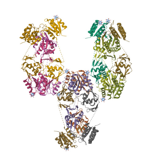
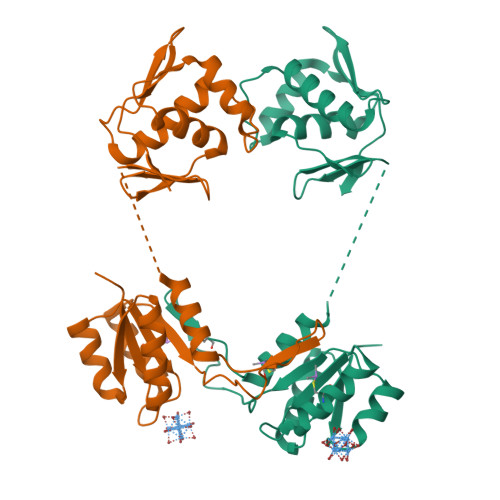
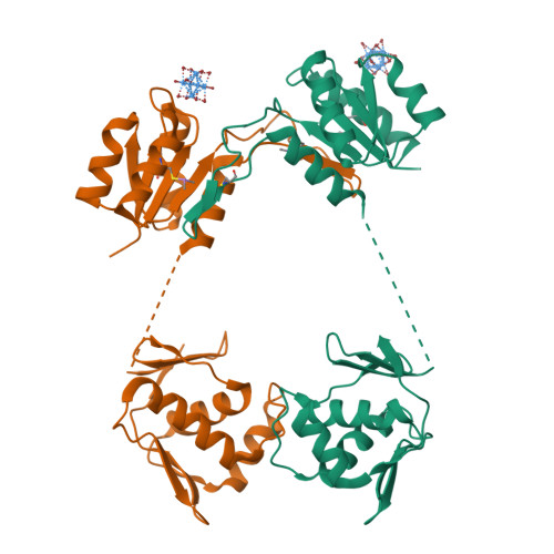
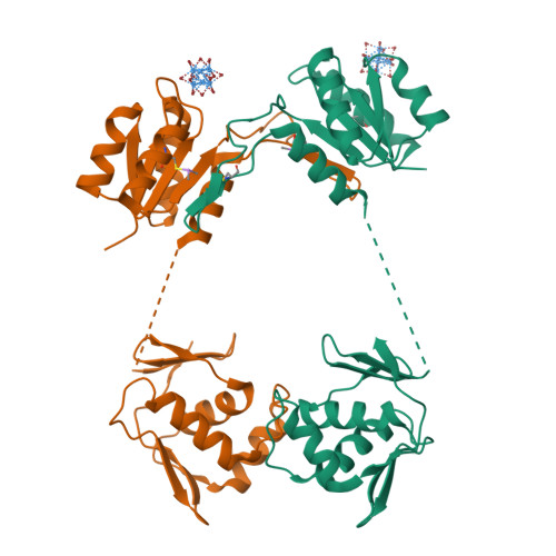
Structure of unphosphorylated BaeR dimer


Experimental Data Snapshot

  • Method: X-RAY DIFFRACTION
  • Resolution: 3.30 Å
  • R-Value Free: 0.245 
  • R-Value Work: 0.225 
  • R-Value Observed: 0.226 

wwPDB Validation   3D Report Full Report


This is version 1.3 of the entry. See complete history


Literature

The Dimeric Form of the Unphosphorylated Response Regulator Baer.

Choudhury, H.G.Beis, K.

(2013) Protein Sci 22: 1287

  • DOI: https://doi.org/10.1002/pro.2311
  • Primary Citation of Related Structures:  
    4B09

  • PubMed Abstract: 

    Bacterial response regulators (RRs) can regulate the expression of genes that confer antibiotic resistance; they contain a receiver and an effector domain and their ability to bind DNA is based on the dimerization state. This is triggered by phosphorylation of the receiver domain by a kinase. However, even in the absence of phosphorylation RRs can exist in equilibrium between monomers and dimers with phosphorylation shifting the equilibrium toward the dimer form. We have determined the crystal structure of the unphosphorylated dimeric BaeR from Escherichia coli. The dimer interface is formed by a domain swap at the receiver domain. In comparison with the unphosphorylated dimeric PhoP from Mycobacterium tuberculosis, BaeR displays an asymmetry of the effector domains.


  • Organizational Affiliation

    Division of Molecular Biosciences, Imperial College London, London, South Kensington, SW7 2AZ, United Kingdom; Membrane Protein Lab, Diamond Light Source, Harwell Science and Innovation Campus, Chilton, Oxfordshire, OX11 0DE, United Kingdom; Research Complex at Harwell, Harwell Oxford, Didcot, Oxforsdhire OX11 0FA, United Kingdom.


Macromolecules
Find similar proteins by:  (by identity cutoff)  |  3D Structure
Entity ID: 1
MoleculeChains Sequence LengthOrganismDetailsImage
TRANSCRIPTIONAL REGULATORY PROTEIN BAER
A, B, C, D, E
A, B, C, D, E, F, G, H, I, J, K, L
240Escherichia coli str. K-12 substr. MG1655Mutation(s): 1 
UniProt
Find proteins for P69229 (Escherichia coli O6:H1 (strain CFT073 / ATCC 700928 / UPEC))
Explore P69229 
Go to UniProtKB:  P69229
Entity Groups  
Sequence Clusters30% Identity50% Identity70% Identity90% Identity95% Identity100% Identity
UniProt GroupP69229
Sequence Annotations
Expand
  • Reference Sequence
Small Molecules
Ligands 1 Unique
IDChains Name / Formula / InChI Key2D Diagram3D Interactions
TBR
Query on TBR

Download Ideal Coordinates CCD File 
M [auth A]
N [auth B]
O [auth C]
P [auth D]
Q [auth E]
M [auth A],
N [auth B],
O [auth C],
P [auth D],
Q [auth E],
R [auth F],
S [auth G],
T [auth I],
U [auth K]
HEXATANTALUM DODECABROMIDE
Br12 Ta6
YWYIQTPPCOBSGN-UHFFFAOYSA-M
Modified Residues  1 Unique
IDChains TypeFormula2D DiagramParent
CAS
Query on CAS
A, B, C, D, E
A, B, C, D, E, F, G, H, I, J, K, L
L-PEPTIDE LINKINGC5 H12 As N O2 SCYS
Experimental Data & Validation

Experimental Data

  • Method: X-RAY DIFFRACTION
  • Resolution: 3.30 Å
  • R-Value Free: 0.245 
  • R-Value Work: 0.225 
  • R-Value Observed: 0.226 
  • Space Group: C 1 2 1
Unit Cell:
Length ( Å )Angle ( ˚ )
a = 236.602α = 90
b = 130.401β = 90.4
c = 198.213γ = 90
Software Package:
Software NamePurpose
XDSdata reduction
SCALAdata scaling
SHELXDphasing
SHARPphasing
PHENIXrefinement

Structure Validation

View Full Validation Report


Entry History 

Deposition Data

Revision History  (Full details and data files)

  • Version 1.0: 2013-07-10
    Type: Initial release
  • Version 1.1: 2013-07-31
    Changes: Database references
  • Version 1.2: 2013-09-25
    Changes: Database references
  • Version 1.3: 2024-11-13
    Changes: Data collection, Database references, Derived calculations, Other, Structure summary