4AZK | pdb_00004azk

Structural basis of L-phosphoserine binding to Bacillus alcalophilus phosphoserine aminotransferase


Experimental Data Snapshot

  • Method: X-RAY DIFFRACTION
  • Resolution: 1.59 Å
  • R-Value Free: 
    0.171 (Depositor), 0.175 (DCC) 
  • R-Value Work: 
    0.153 (Depositor), 0.158 (DCC) 
  • R-Value Observed: 
    0.154 (Depositor) 

Starting Model: experimental
View more details

wwPDB Validation 3D Report Full Report

Validation slider image for 4AZK

Ligand Structure Quality Assessment 


This is version 1.3 of the entry. See complete history

Literature

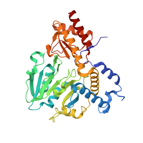
Structural Basis of L-Phosphoserine Binding to Bacillus Alcalophilus Phosphoserine Aminotransferase

Battula, P.Dubnovitsky, A.P.Papageorgiou, A.C.

(2013) Acta Crystallogr D Biol Crystallogr 69: 804

  • DOI: https://doi.org/10.1107/S0907444913002096
  • Primary Citation Related Structures: 
    4AZJ, 4AZK

  • PubMed Abstract: 

    Phosphoserine aminotransferase is a vitamin B6-dependent enzyme that catalyzes the reversible conversion of 3-phosphohydroxypyruvate to L-phosphoserine using glutamate as an amine donor. In an effort to gain insight into the substrate-recognition mechanism of the enzyme, crystal structures of Bacillus alcalophilus phosphoserine aminotransferase in the presence or absence of L-phosphoserine were determined to resolutions of 1.5 and 1.6 Å, respectively. Local conformational changes induced upon substrate binding were identified. However, in contrast to other aminotransferases, no domain or subunit movements were observed. Two Arg residues (Arg42 and Arg328) and two His residues (His41 and His327) were found to form a tight binding site for the phosphate group of L-phosphoserine. Comparison with Escherichia coli phosphoserine aminotransferase in complex with the substrate analogue α-methylglutamate revealed more extensive structural changes in the case of L-phosphoserine binding. Based on the structural analysis, the flexibility of Arg328 is proposed to be critical for substrate recognition.


  • Organizational Affiliation
    • Turku Centre for Biotechnology, University of Turku and Åbo Akademi University, BioCity, FIN-20521 Turku, Finland.

Macromolecule Content 

  • Total Structure Weight: 81.05 kDa 
  • Atom Count: 6,868 
  • Modeled Residue Count: 718 
  • Deposited Residue Count: 720 
  • Unique protein chains: 1

Macromolecules

Find similar proteins by:|  3D Structure
Entity ID: 1
MoleculeChains  Sequence LengthOrganismDetailsImage
PHOSPHOSERINE AMINOTRANSFERASE
A, B
360Alkalihalobacillus alcalophilusMutation(s): 0 
EC: 2.6.1.52
UniProt
Find proteins for Q9RME2 (Alkalihalobacillus alcalophilus)
Explore Q9RME2 
Go to UniProtKB:  Q9RME2
Entity Groups
Sequence Clusters30% Identity50% Identity70% Identity90% Identity95% Identity100% Identity
UniProt GroupQ9RME2
Sequence Annotations
Expand
Reference Sequence

Experimental Data & Validation

Experimental Data

  • Method: X-RAY DIFFRACTION
  • Resolution: 1.59 Å
  • R-Value Free:  0.171 (Depositor), 0.175 (DCC) 
  • R-Value Work:  0.153 (Depositor), 0.158 (DCC) 
  • R-Value Observed: 0.154 (Depositor) 
Space Group: C 2 2 21
Unit Cell:
Length ( Å )Angle ( ˚ )
a = 105.598α = 90
b = 136.599β = 90
c = 152.004γ = 90
Software Package:
Software NamePurpose
PHENIXrefinement
DENZOdata reduction
SCALEPACKdata scaling

Structure Validation

View Full Validation Report


Ligand Structure Quality Assessment 


Entry History 

Deposition Data

Revision History  (Full details and data files)

  • Version 1.0: 2013-05-01
    Type: Initial release
  • Version 1.1: 2013-05-15
    Changes: Database references
  • Version 1.2: 2019-07-17
    Changes: Data collection, Derived calculations
  • Version 1.3: 2023-12-20
    Changes: Data collection, Database references, Derived calculations, Other, Refinement description