Design and Evaluation of Meningococcal Vaccines Through Structure-Based Modification of Host and Pathogen Molecules
Johnson, S., Tan, L., Van Der Veen, S., Caesar, J., Goicoechea De Jorge, E., Harding, R.J., Bai, X., Exley, R.M., Ward, P.N., Ruivo, N., Trivedi, K., Cumber, E., Jones, R., Newham, L., Staunton, D., Ufret-Vincenty, R., Borrow, R., Pickering, M., Lea, S.M., Tang, C.M.(2012) PLoS Pathog 8: 2981
- PubMed: 23133374
- DOI: https://doi.org/10.1371/journal.ppat.1002981
- Primary Citation of Related Structures:
2YBY, 4AYD, 4AYE, 4AYI, 4AYM, 4AYN - PubMed Abstract:
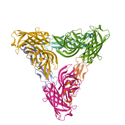
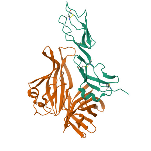
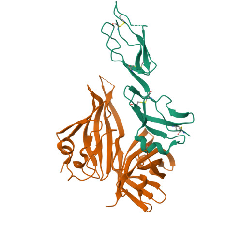
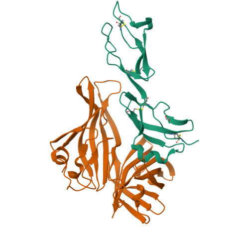
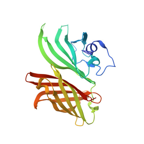
Neisseria meningitis remains a leading cause of sepsis and meningitis, and vaccines are required to prevent infections by this important human pathogen. Factor H binding protein (fHbp) is a key antigen that elicits protective immunity against the meningococcus and recruits the host complement regulator, fH. As the high affinity interaction between fHbp and fH could impair immune responses, we sought to identify non-functional fHbps that could act as effective immunogens. This was achieved by alanine substitution of fHbps from all three variant groups (V1, V2 and V3 fHbp) of the protein; while some residues affected fH binding in each variant group, the distribution of key amino underlying the interaction with fH differed between the V1, V2 and V3 proteins. The atomic structure of V3 fHbp in complex with fH and of the C-terminal barrel of V2 fHbp provide explanations to the differences in the precise nature of their interactions with fH, and the instability of the V2 protein. To develop transgenic models to assess the efficacy of non-functional fHbps, we determined the structural basis of the low level of interaction between fHbp and murine fH; in addition to changes in amino acids in the fHbp binding site, murine fH has a distinct conformation compared with the human protein that would sterically inhibit binding to fHbp. Non-functional V1 fHbps were further characterised by binding and structural studies, and shown in non-transgenic and transgenic mice (expressing chimeric fH that binds fHbp and precisely regulates complement system) to retain their immunogenicity. Our findings provide a catalogue of non-functional fHbps from all variant groups that can be included in new generation meningococcal vaccines, and establish proof-in-principle for clinical studies to compare their efficacy with wild-type fHbps.
Organizational Affiliation:
Sir William Dunn School of Pathology, University of Oxford, Oxford, United Kingdom.


