4AW4 | pdb_00004aw4

Engineered variant of Listeria monocytogenes InlB internalin domain with an additional leucine rich repeat inserted


Experimental Data Snapshot

  • Method: X-RAY DIFFRACTION
  • Resolution: 1.93 Å
  • R-Value Free: 
    0.205 (Depositor), 0.209 (DCC) 
  • R-Value Work: 
    0.167 (Depositor), 0.172 (DCC) 
  • R-Value Observed: 
    0.168 (Depositor) 

Starting Model: experimental
View more details

wwPDB Validation 3D Report Full Report

Validation slider image for 4AW4

This is version 1.6 of the entry. See complete history

Literature

Engineered Variants of Inlb with an Additional Leucine-Rich Repeat Discriminate between Physiologically Relevant and Packing Contacts in Crystal Structures of the Inlb:Met Complex.

Niemann, H.H.Gherardi, E.Bleymuller, W.M.Heinz, D.W.

(2012) Protein Sci 21: 1528

  • DOI: https://doi.org/10.1002/pro.2142
  • Primary Citation Related Structures: 
    4AW4

  • PubMed Abstract: 

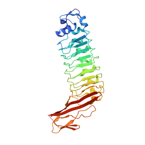
    The physiological relevance of contacts in crystal lattices often remains elusive. This was also the case for the complex between the invasion protein internalin B (InlB) from Listeria monocytogenes and its host cell receptor, the human receptor tyrosine kinase (RTK) MET. InlB is a MET agonist and induces bacterial host cell invasion. Activation of RTKs generally involves ligand-induced dimerization of the receptor ectodomain. The two currently available crystal structures of the InlB:MET complex show the same arrangement of InlB and MET in a 1:1 complex, but different dimeric 2:2 assemblies. Only one of these 2:2 assemblies is predicted to be stable by a computational procedure. This assembly is mainly stabilized by a contact between the Cap domain of InlB from one and the Sema domain of MET from another 1:1 complex. Here, we probe the physiological relevance of this interaction. We generated variants of the leucine-rich repeat (LRR) protein InlB by inserting an additional repeat between the first and the second LRR. This should allow formation of the 1:1 complex but disrupt the potential 2:2 complex involving the Cap-Sema contact due to steric distortions. A crystal structure of one of the engineered proteins showed that it folded properly. Binding affinity to MET was comparable to that of wild-type InlB. The InlB variant induced MET phosphorylation and cell scatter like wild-type InlB. These results suggest that the Cap-Sema interaction is not physiologically relevant and support the previously proposed assembly, in which a 2:2 InlB:MET complex is built around a ligand dimer.


  • Organizational Affiliation
    • Department of Chemistry, Bielefeld University, 33501 Bielefeld, Germany. Hartmut.Niemann@uni-bielefeld.de

Macromolecule Content 

  • Total Structure Weight: 107.18 kDa 
  • Atom Count: 8,080 
  • Modeled Residue Count: 915 
  • Deposited Residue Count: 933 
  • Unique protein chains: 1

Macromolecules

Find similar proteins by:|  3D Structure
Entity ID: 1
MoleculeChains  Sequence LengthOrganismDetailsImage
INTERNALIN B
A, B, C
311Listeria monocytogenes EGD-eMutation(s): 0 
UniProt
Find proteins for P0DQD3 (Listeria monocytogenes serotype 1/2a (strain EGD / Mackaness))
Explore P0DQD3 
Go to UniProtKB:  P0DQD3
Entity Groups
Sequence Clusters30% Identity50% Identity70% Identity90% Identity95% Identity100% Identity
UniProt GroupP0DQD3
Sequence Annotations
Expand
Reference Sequence

Small Molecules

Ligands 2 Unique
IDChains Name / Formula / InChI Key2D Diagram3D Interactions
SO4

Query on SO4


Download:Ideal Coordinates CCD File
AA [auth B]
BA [auth B]
CA [auth C]
D [auth A]
E [auth A]
AA [auth B],
BA [auth B],
CA [auth C],
D [auth A],
E [auth A],
F [auth A],
FA [auth C],
G [auth A],
GA [auth C],
H [auth A],
HA [auth C],
I [auth A],
IA [auth C],
JA [auth C],
O [auth A],
P [auth A],
Q [auth A],
S [auth B],
T [auth B],
X [auth B],
Y [auth B],
Z [auth B]
SULFATE ION
O4 S
QAOWNCQODCNURD-UHFFFAOYSA-L
GOL

Query on GOL


Download:Ideal Coordinates CCD File
DA [auth C]
EA [auth C]
J [auth A]
K [auth A]
L [auth A]
DA [auth C],
EA [auth C],
J [auth A],
K [auth A],
L [auth A],
M [auth A],
N [auth A],
R [auth B],
U [auth B],
V [auth B],
W [auth B]
GLYCEROL
C3 H8 O3
PEDCQBHIVMGVHV-UHFFFAOYSA-N

Experimental Data & Validation

Experimental Data

  • Method: X-RAY DIFFRACTION
  • Resolution: 1.93 Å
  • R-Value Free:  0.205 (Depositor), 0.209 (DCC) 
  • R-Value Work:  0.167 (Depositor), 0.172 (DCC) 
  • R-Value Observed: 0.168 (Depositor) 
Space Group: C 1 2 1
Unit Cell:
Length ( Å )Angle ( ˚ )
a = 179.08α = 90
b = 102.15β = 90.23
c = 69.81γ = 90
Software Package:
Software NamePurpose
REFMACrefinement
XDSdata reduction
XSCALEdata scaling
PHASERphasing

Structure Validation

View Full Validation Report


Entry History 

Deposition Data

Revision History  (Full details and data files)

  • Version 1.0: 2012-08-15
    Type: Initial release
  • Version 1.1: 2012-08-29
    Changes: Database references
  • Version 1.2: 2012-10-03
    Changes: Database references
  • Version 1.3: 2019-05-08
    Changes: Data collection, Derived calculations, Experimental preparation, Other
  • Version 1.4: 2019-05-22
    Changes: Data collection, Experimental preparation
  • Version 1.5: 2019-07-17
    Changes: Data collection
  • Version 1.6: 2023-12-20
    Changes: Data collection, Database references, Derived calculations, Other, Refinement description