4ASX | pdb_00004asx

Crystal structure of Activin receptor type-IIA (ACVR2A) kinase domain in complex with dihydro-Bauerine C


Experimental Data Snapshot

  • Method: X-RAY DIFFRACTION
  • Resolution: 2.05 Å
  • R-Value Free: 
    0.225 (Depositor), 0.224 (DCC) 
  • R-Value Work: 
    0.187 (Depositor), 0.189 (DCC) 
  • R-Value Observed: 
    0.188 (Depositor) 

Starting Model: experimental
View more details

wwPDB Validation 3D Report Full Report

Validation slider image for 4ASX

Ligand Structure Quality Assessment 


This is version 1.4 of the entry. See complete history

Literature

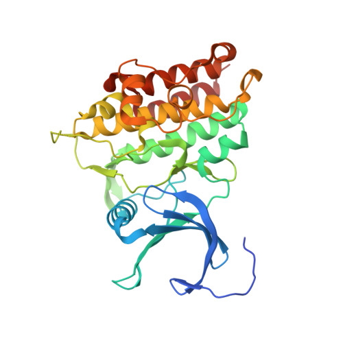
Crystal Structure of Activin Receptor Type-Iia (Acvr2A) Kinase Domain in Complex with a Beta- Carboline Inhibitor

Williams, E.Chaikuad, A.Canning, P.Mahajan, P.Cooper, C.D.O.Beltrami, A.Krojer, T.Huber, K.Bracher, F.von Delft, F.Arrowsmith, C.H.Edwards, A.M.Bountra, C.Bullock, A.

To be published.

Macromolecule Content 

  • Total Structure Weight: 74.19 kDa 
  • Atom Count: 5,328 
  • Modeled Residue Count: 617 
  • Deposited Residue Count: 644 
  • Unique protein chains: 1

Macromolecules

Find similar proteins by:|  3D Structure
Entity ID: 1
MoleculeChains  Sequence LengthOrganismDetailsImage
ACTIVIN RECEPTOR TYPE-2A
A, B
322Homo sapiensMutation(s): 0 
EC: 2.7.11.30
UniProt & NIH Common Fund Data Resources
Find proteins for P27037 (Homo sapiens)
Explore P27037 
Go to UniProtKB:  P27037
PHAROS:  P27037
GTEx:  ENSG00000121989 
Entity Groups
Sequence Clusters30% Identity50% Identity70% Identity90% Identity95% Identity100% Identity
UniProt GroupP27037
Sequence Annotations
Expand
Reference Sequence

Small Molecules

Ligands 2 Unique
IDChains Name / Formula / InChI Key2D Diagram3D Interactions
6OJ

Query on 6OJ


Download:Ideal Coordinates CCD File
C [auth A],
H [auth B]
7,8-bis(chloranyl)-9-methyl-3,4-dihydro-2H-pyrido[3,4-b]indol-1-one
C12 H10 Cl2 N2 O
MYIBLDZPAOEFTB-UHFFFAOYSA-N
EDO

Query on EDO


Download:Ideal Coordinates CCD File
D [auth A]
E [auth A]
F [auth A]
G [auth A]
I [auth B]
D [auth A],
E [auth A],
F [auth A],
G [auth A],
I [auth B],
J [auth B],
K [auth B],
L [auth B],
M [auth B]
1,2-ETHANEDIOL
C2 H6 O2
LYCAIKOWRPUZTN-UHFFFAOYSA-N

Experimental Data & Validation

Experimental Data

  • Method: X-RAY DIFFRACTION
  • Resolution: 2.05 Å
  • R-Value Free:  0.225 (Depositor), 0.224 (DCC) 
  • R-Value Work:  0.187 (Depositor), 0.189 (DCC) 
  • R-Value Observed: 0.188 (Depositor) 
Space Group: P 65 2 2
Unit Cell:
Length ( Å )Angle ( ˚ )
a = 110.426α = 90
b = 110.426β = 90
c = 208.848γ = 120
Software Package:
Software NamePurpose
REFMACrefinement
XDSdata reduction
SCALAdata scaling
PHASERphasing

Structure Validation

View Full Validation Report


Ligand Structure Quality Assessment 


Entry History 

Revision History  (Full details and data files)

  • Version 1.0: 2012-05-23
    Type: Initial release
  • Version 1.1: 2012-12-05
    Changes: Database references, Structure summary
  • Version 1.2: 2018-01-24
    Changes: Database references, Structure summary
  • Version 1.3: 2018-06-20
    Changes: Data collection, Derived calculations
  • Version 1.4: 2023-12-20
    Changes: Data collection, Database references, Derived calculations, Other, Refinement description