4AS8 | pdb_00004as8

X-ray structure of the cyan fluorescent protein Cerulean cryoprotected with ethylene glycol


Experimental Data Snapshot

  • Method: X-RAY DIFFRACTION
  • Resolution: 1.02 Å
  • R-Value Free: 
    0.132 (Depositor), 0.142 (DCC) 
  • R-Value Work: 
    0.116 (Depositor), 0.128 (DCC) 
  • R-Value Observed: 
    0.117 (Depositor) 

Starting Model: experimental
View more details

wwPDB Validation 3D Report Full Report

Validation slider image for 4AS8

This is version 1.6 of the entry. See complete history

Literature

Alteration of Fluorescent Protein Spectroscopic Properties Upon Cryoprotection

von Stetten, D.Batot, G.Noirclerc-Savoye, M.Royant, A.

(2012) Acta Crystallogr D Biol Crystallogr 68: 1578

  • DOI: https://doi.org/10.1107/S0907444912037900
  • Primary Citation Related Structures: 
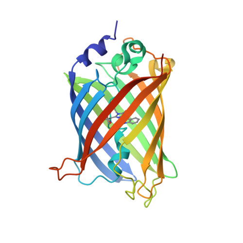
    4AS8

  • PubMed Abstract: 

    Cryoprotection of a protein crystal by addition of small-molecule compounds may sometimes affect the structure of its active site. The spectroscopic and structural effects of the two cryoprotectants glycerol and ethylene glycol on the cyan fluorescent protein Cerulean were investigated. While glycerol had almost no noticeable effect, ethylene glycol was shown to induce a systematic red shift of the UV-vis absorption and fluorescence emission spectra. Additionally, ethylene glycol molecules were shown to enter the core of the protein, with one of them binding in close vicinity to the chromophore, which provides a sound explanation for the observed spectroscopic changes. These results highlight the need to systematically record spectroscopic data on crystals of light-absorbing proteins and reinforce the notion that fluorescent proteins must not been seen as rigid structures.


  • Organizational Affiliation
    • Structural Biology Group, European Synchrotron Radiation Facility, Grenoble, France. vonstett@esrf.fr

Macromolecule Content 

  • Total Structure Weight: 28.33 kDa 
  • Atom Count: 2,303 
  • Modeled Residue Count: 229 
  • Deposited Residue Count: 243 
  • Unique protein chains: 1

Macromolecules

Find similar proteins by:|  3D Structure
Entity ID: 1
MoleculeChains  Sequence LengthOrganismDetailsImage
GREEN FLUORESCENT PROTEIN243Aequorea victoriaMutation(s): 8 
UniProt
Find proteins for P42212 (Aequorea victoria)
Explore P42212 
Go to UniProtKB:  P42212
Entity Groups
Sequence Clusters30% Identity50% Identity70% Identity90% Identity95% Identity100% Identity
UniProt GroupP42212
Sequence Annotations
Expand
Reference Sequence

Small Molecules

Ligands 2 Unique
IDChains Name / Formula / InChI Key2D Diagram3D Interactions
EDO

Query on EDO


Download:Ideal Coordinates CCD File
B [auth A]
C [auth A]
D [auth A]
E [auth A]
F [auth A]
B [auth A],
C [auth A],
D [auth A],
E [auth A],
F [auth A],
G [auth A],
H [auth A],
I [auth A],
J [auth A],
K [auth A],
L [auth A]
1,2-ETHANEDIOL
C2 H6 O2
LYCAIKOWRPUZTN-UHFFFAOYSA-N
MG

Query on MG


Download:Ideal Coordinates CCD File
M [auth A]MAGNESIUM ION
Mg
JLVVSXFLKOJNIY-UHFFFAOYSA-N
Modified Residues  1 Unique
IDChains TypeFormula2D DiagramParent
CRF
Query on CRF
A
L-PEPTIDE LINKINGC17 H18 N4 O4THR, TRP, GLY

Experimental Data & Validation

Experimental Data

  • Method: X-RAY DIFFRACTION
  • Resolution: 1.02 Å
  • R-Value Free:  0.132 (Depositor), 0.142 (DCC) 
  • R-Value Work:  0.116 (Depositor), 0.128 (DCC) 
  • R-Value Observed: 0.117 (Depositor) 
Space Group: P 21 21 21
Unit Cell:
Length ( Å )Angle ( ˚ )
a = 51.344α = 90
b = 62.662β = 90
c = 70.205γ = 90
Software Package:
Software NamePurpose
REFMACrefinement
XDSdata reduction
SCALAdata scaling
REFMACphasing

Structure Validation

View Full Validation Report


Entry History 

Deposition Data

Revision History  (Full details and data files)

  • Version 1.0: 2012-10-31
    Type: Initial release
  • Version 1.1: 2012-11-07
    Changes: Database references
  • Version 1.2: 2017-09-27
    Changes: Data collection
  • Version 1.3: 2017-12-20
    Changes: Database references
  • Version 1.4: 2019-10-23
    Changes: Data collection, Database references, Derived calculations, Other
  • Version 1.5: 2023-12-20
    Changes: Data collection, Database references, Derived calculations, Refinement description
  • Version 1.6: 2024-11-13
    Changes: Structure summary