4AQ5 | pdb_00004aq5

Gating movement in acetylcholine receptor analysed by time-resolved electron cryo-microscopy (closed class)


Experimental Data Snapshot

  • Method: ELECTRON MICROSCOPY
  • Resolution: 6.20 Å
  • Aggregation State: HELICAL ARRAY 
  • Reconstruction Method: HELICAL 

Starting Model: experimental
View more details

wwPDB Validation 3D Report Full Report

Validation slider image for 4AQ5

This is version 1.5 of the entry. See complete history

Literature

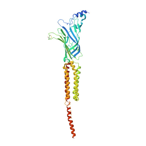
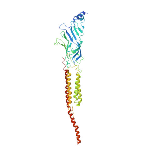
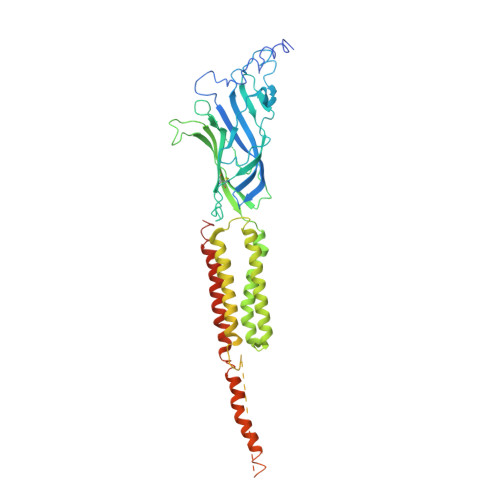
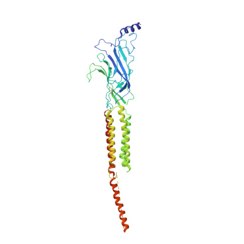
Gating Movement of Acetylcholine Receptor Caught by Plunge-Freezing.

Unwin, N.Fujiyoshi, Y.

(2012) J Mol Biology 422: 617

  • DOI: https://doi.org/10.1016/j.jmb.2012.07.010
  • Primary Citation Related Structures: 
    4AQ5, 4AQ9

  • PubMed Abstract: 

    The nicotinic acetylcholine (ACh) receptor converts transiently to an open-channel form when activated by ACh released into the synaptic cleft. We describe here the conformational change underlying this event, determined by electron microscopy of ACh-sprayed and freeze-trapped postsynaptic membranes. ACh binding to the α subunits triggers a concerted rearrangement in the ligand-binding domain, involving an ~1-Å outward displacement of the extracellular portion of the β subunit where it interacts with the juxtaposed ends of α-helices shaping the narrow membrane-spanning pore. The β-subunit helices tilt outward to accommodate this displacement, destabilising the arrangement of pore-lining helices, which in the closed channel bend inward symmetrically to form a central hydrophobic gate. Straightening and tangential motion of the pore-lining helices effect channel opening by widening the pore asymmetrically and increasing its polarity in the region of the gate. The pore-lining helices of the α(γ) and δ subunits, by flexing between alternative bent and straight conformations, undergo the greatest movements. This coupled allosteric transition shifts the structure from a tense (closed) state toward a more relaxed (open) state.


  • Organizational Affiliation
    • MRC Laboratory of Molecular Biology, Hills Road, Cambridge CB2 0QH, UK. Electronic address: unwin@mrc-lmb.cam.ac.uk.

Macromolecule Content 

  • Total Structure Weight: 278.07 kDa 
  • Atom Count: 14,924 
  • Modeled Residue Count: 1,851 
  • Deposited Residue Count: 2,425 
  • Unique protein chains: 4

Macromolecules

Find similar proteins by:|  3D Structure
Entity ID: 1
MoleculeChains  Sequence LengthOrganismDetailsImage
ACETYLCHOLINE RECEPTOR SUBUNIT ALPHA
A, D
461Torpedo marmorataMutation(s): 0 
Membrane Entity: Yes 
UniProt
Find proteins for P02711 (Torpedo marmorata)
Explore P02711 
Go to UniProtKB:  P02711
Entity Groups
Sequence Clusters30% Identity50% Identity70% Identity90% Identity95% Identity100% Identity
UniProt GroupP02711
Sequence Annotations
Expand
Reference Sequence
Find similar proteins by:|  3D Structure
Entity ID: 2
MoleculeChains  Sequence LengthOrganismDetailsImage
ACETYLCHOLINE RECEPTOR BETA SUBUNIT493Torpedo marmorataMutation(s): 0 
Membrane Entity: Yes 
UniProt
Find proteins for Q6S3I0 (Torpedo marmorata)
Explore Q6S3I0 
Go to UniProtKB:  Q6S3I0
Entity Groups
Sequence Clusters30% Identity50% Identity70% Identity90% Identity95% Identity100% Identity
UniProt GroupQ6S3I0
Sequence Annotations
Expand
Reference Sequence
Find similar proteins by:|  3D Structure
Entity ID: 3
MoleculeChains  Sequence LengthOrganismDetailsImage
ACETYLCHOLINE RECEPTOR DELTA SUBUNIT522Torpedo marmorataMutation(s): 0 
Membrane Entity: Yes 
UniProt
Find proteins for Q6S3H8 (Torpedo marmorata)
Explore Q6S3H8 
Go to UniProtKB:  Q6S3H8
Entity Groups
Sequence Clusters30% Identity50% Identity70% Identity90% Identity95% Identity100% Identity
UniProt GroupQ6S3H8
Sequence Annotations
Expand
Reference Sequence
Find similar proteins by:|  3D Structure
Entity ID: 4
MoleculeChains  Sequence LengthOrganismDetailsImage
ACETYLCHOLINE RECEPTOR GAMMA SUBUNIT488Torpedo marmorataMutation(s): 0 
Membrane Entity: Yes 
UniProt
Find proteins for Q6S3H9 (Torpedo marmorata)
Explore Q6S3H9 
Go to UniProtKB:  Q6S3H9
Entity Groups
Sequence Clusters30% Identity50% Identity70% Identity90% Identity95% Identity100% Identity
UniProt GroupQ6S3H9
Sequence Annotations
Expand
Reference Sequence

Experimental Data & Validation

Experimental Data

  • Method: ELECTRON MICROSCOPY
  • Resolution: 6.20 Å
  • Aggregation State: HELICAL ARRAY 
  • Reconstruction Method: HELICAL 
EM Software:
TaskSoftware PackageVersion
RECONSTRUCTIONCustom
RECONSTRUCTIONMRC IMAGE PROCESSING PACKAGE

Structure Validation

View Full Validation Report


Entry History 

Deposition Data

Revision History  (Full details and data files)

  • Version 1.0: 2012-08-01
    Type: Initial release
  • Version 1.1: 2012-09-12
    Changes: Database references
  • Version 1.2: 2013-09-25
    Changes: Other
  • Version 1.3: 2017-08-30
    Changes: Data collection
  • Version 1.4: 2019-10-23
    Changes: Data collection, Other
  • Version 1.5: 2024-11-20
    Changes: Data collection, Database references, Refinement description, Structure summary