4APR | pdb_00004apr

STRUCTURES OF COMPLEXES OF RHIZOPUSPEPSIN WITH PEPSTATIN AND OTHER STATINE-CONTAINING INHIBITORS


Experimental Data Snapshot

  • Method: X-RAY DIFFRACTION
  • Resolution: 2.50 Å
  • R-Value Work: 
    0.146 (DCC) 
  • R-Value Observed: 
    0.154 (Depositor) 

wwPDB Validation 3D Report Full Report

Validation slider image for 4APR

This is version 1.5 of the entry. See complete history

Literature

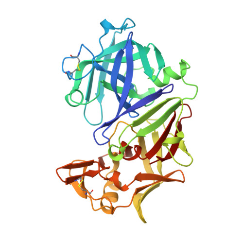
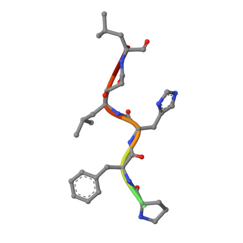
Structures of complexes of rhizopuspepsin with pepstatin and other statine-containing inhibitors.

Suguna, K.Padlan, E.A.Bott, R.Boger, J.Parris, K.D.Davies, D.R.

(1992) Proteins 13: 195-205

  • DOI: https://doi.org/10.1002/prot.340130303
  • Primary Citation Related Structures: 
    4APR, 5APR, 6APR

  • PubMed Abstract: 

    The three-dimensional structures of the complexes of the aspartic proteinase from Rhizopus chinensis (Rhizopuspepsin, EC 3.4.23.6) with pepstatin and two pepstatin-like peptide inhibitors of renin have been determined by X-ray diffraction methods and refined by restrained least-squares procedures. The inhibitors adopt an extended conformation and lie in the deep groove located between the two domains of the enzyme. Inhibitor binding is accompanied by a conformational change at the "flap," a beta-hairpin loop region, that projects over the binding cleft and closes down over the inhibitor, excluding water molecules from the vicinity of the scissile bond. The hydroxyl group of the central statyl residue of the inhibitors replaces the water molecule found between the two active aspartates, Asp-35 and Asp-218, in the native structure. The refined structures provide additional data to define the specific subsites of the enzyme and also show a system of hydrogen bonding to the inhibitor backbone similar to that observed for a reduced inhibitor.


  • Organizational Affiliation
    • Laboratory of Molecular Biology, National Institute of Diabetes and Digestive and Kidney Diseases, National Institutes of Health, Bethesda, Maryland 20892.

Macromolecule Content 

  • Total Structure Weight: 35.11 kDa 
  • Atom Count: 2,776 
  • Modeled Residue Count: 330 
  • Deposited Residue Count: 333 
  • Unique protein chains: 2

Macromolecules

Find similar proteins by:|  3D Structure
Entity ID: 1
MoleculeChains  Sequence LengthOrganismDetailsImage
RHIZOPUSPEPSINA [auth E]325Rhizopus microsporus var. chinensisMutation(s): 0 
EC: 3.4.23.6 (PDB Primary Data), 3.4.23.21 (UniProt)
UniProt
Find proteins for P06026 (Rhizopus chinensis)
Explore P06026 
Go to UniProtKB:  P06026
Entity Groups
Sequence Clusters30% Identity50% Identity70% Identity90% Identity95% Identity100% Identity
UniProt GroupP06026
Sequence Annotations
Expand
Reference Sequence
Find similar proteins by:  Sequence   |   3D Structure  
Entity ID: 2
MoleculeChains  Sequence LengthOrganismDetailsImage
PEPSTATIN-LIKE RENIN INHIBITORB [auth I]8N/AMutation(s): 0 

Experimental Data & Validation

Experimental Data

  • Method: X-RAY DIFFRACTION
  • Resolution: 2.50 Å
  • R-Value Work:  0.146 (DCC) 
  • R-Value Observed: 0.154 (Depositor) 
Space Group: P 21 21 21
Unit Cell:
Length ( Å )Angle ( ˚ )
a = 60.39α = 90
b = 60.61β = 90
c = 106.9γ = 90
Software Package:
Software NamePurpose
PROLSQrefinement

Structure Validation

View Full Validation Report


Entry History 

Deposition Data

Revision History  (Full details and data files)

  • Version 1.0: 1991-04-15
    Type: Initial release
  • Version 1.1: 2008-03-03
    Changes: Version format compliance
  • Version 1.2: 2011-07-13
    Changes: Atomic model, Database references, Derived calculations, Non-polymer description, Structure summary, Version format compliance
  • Version 1.3: 2012-05-23
    Changes: Source and taxonomy
  • Version 1.4: 2017-11-29
    Changes: Derived calculations, Other
  • Version 1.5: 2024-10-23
    Changes: Data collection, Database references, Derived calculations, Structure summary