4AMV | pdb_00004amv

E.COLI GLUCOSAMINE-6P SYNTHASE IN COMPLEX WITH FRUCTOSE-6P


Experimental Data Snapshot

  • Method: X-RAY DIFFRACTION
  • Resolution: 2.05 Å
  • R-Value Free: 
    0.240 (Depositor), 0.238 (DCC) 
  • R-Value Work: 
    0.206 (Depositor), 0.205 (DCC) 
  • R-Value Observed: 
    0.208 (Depositor) 

Starting Model: experimental
View more details

wwPDB Validation 3D Report Full Report

Validation slider image for 4AMV

Ligand Structure Quality Assessment 


This is version 1.1 of the entry. See complete history

Literature

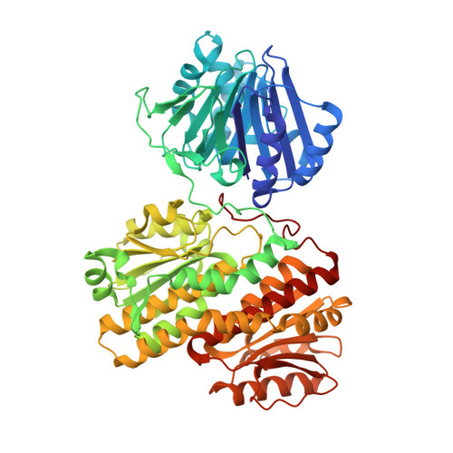
Glutamine Binding Opens the Ammonia Channel and Activates Glucosamine-6P Synthase

Mouilleron, S.Badet-Denisot, M.A.Golinelli-Pimpaneau, B.

(2006) J Biological Chem 281: 4404

  • DOI: https://doi.org/10.1074/jbc.M511689200
  • Primary Citation Related Structures: 
    2J6H, 4AMV

  • PubMed Abstract: 

    Glucosamine-6P synthase catalyzes the synthesis of glucosamine-6P from fructose-6P and glutamine and uses a channel to transfer ammonia from its glutaminase to its synthase active site. X-ray structures of glucosamine-6P synthase have been determined at 2.05 Angstroms resolution in the presence of fructose-6P and at 2.35 Angstroms resolution in the presence of fructose-6P and 6-diazo-5-oxo-L-norleucine, a glutamine affinity analog that covalently modifies the N-terminal catalytic cysteine, therefore mimicking the gamma-glutamyl-thioester intermediate formed during hydrolysis of glutamine. The fixation of the glutamine analog activates the enzyme through several major structural changes: 1) the closure of a loop to shield the glutaminase site accompanied by significant domain hinging, 2) the activation of catalytic residues involved in glutamine hydrolysis, i.e. the alpha-amino group of Cys-1 and Asn-98 that is positioned to form the oxyanion hole, and 3) a 75 degrees rotation of the Trp-74 indole group that opens the ammonia channel.


  • Organizational Affiliation
    • Laboratoire d'Enzymologie et de Biochimie Structurales, Unite Propre de Recherche 9063, France.

Macromolecule Content 

  • Total Structure Weight: 201.63 kDa 
  • Atom Count: 14,225 
  • Modeled Residue Count: 1,824 
  • Deposited Residue Count: 1,827 
  • Unique protein chains: 1

Macromolecules

Find similar proteins by:|  3D Structure
Entity ID: 1
MoleculeChains  Sequence LengthOrganismDetailsImage
GLUCOSAMINE--FRUCTOSE-6-PHOSPHATE AMINOTRANSFERASE [ISOMER IZING]
A, B, C
609Escherichia coliMutation(s): 0 
EC: 2.6.1.16
UniProt
Find proteins for P17169 (Escherichia coli (strain K12))
Explore P17169 
Go to UniProtKB:  P17169
Entity Groups
Sequence Clusters30% Identity50% Identity70% Identity90% Identity95% Identity100% Identity
UniProt GroupP17169
Sequence Annotations
Expand
Reference Sequence

Experimental Data & Validation

Experimental Data

  • Method: X-RAY DIFFRACTION
  • Resolution: 2.05 Å
  • R-Value Free:  0.240 (Depositor), 0.238 (DCC) 
  • R-Value Work:  0.206 (Depositor), 0.205 (DCC) 
  • R-Value Observed: 0.208 (Depositor) 
Space Group: C 1 2 1
Unit Cell:
Length ( Å )Angle ( ˚ )
a = 132.197α = 90
b = 109.677β = 97.11
c = 176.332γ = 90
Software Package:
Software NamePurpose
REFMACrefinement
DENZOdata reduction
SCALEPACKdata scaling

Structure Validation

View Full Validation Report


Ligand Structure Quality Assessment 


Entry History 

Deposition Data

Revision History  (Full details and data files)

  • Version 1.0: 2013-04-24
    Type: Initial release
  • Version 1.1: 2023-12-20
    Changes: Data collection, Database references, Derived calculations, Other, Refinement description