4AMH | pdb_00004amh

Influence of circular permutation on the folding pathway of a PDZ domain


Experimental Data Snapshot

  • Method: X-RAY DIFFRACTION
  • Resolution: 2.30 Å
  • R-Value Free: 
    0.268 (Depositor), 0.273 (DCC) 
  • R-Value Work: 
    0.220 (Depositor), 0.216 (DCC) 
  • R-Value Observed: 
    0.222 (Depositor) 

Starting Model: experimental
View more details

wwPDB Validation 3D Report Full Report

Validation slider image for 4AMH

This is version 1.3 of the entry. See complete history

Literature

Tolerance of Protein Folding to a Circular Permutation in a Pdz Domain

Hultqvist, G.Punekar, A.S.Morrone, A.Chi, C.N.Engstrom, A.Selmer, M.Gianni, S.Jemth, P.

(2012) PLoS One 7: 50055

  • DOI: https://doi.org/10.1371/journal.pone.0050055
  • Primary Citation Related Structures: 
    4AMH

  • PubMed Abstract: 

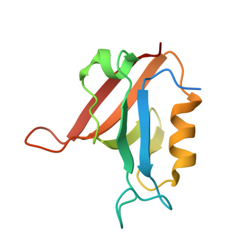
    Circular permutation is a common molecular mechanism for evolution of proteins. However, such re-arrangement of secondary structure connectivity may interfere with the folding mechanism causing accumulation of folding intermediates, which in turn can lead to misfolding. We solved the crystal structure and investigated the folding pathway of a circularly permuted variant of a PDZ domain, SAP97 PDZ2. Our data illustrate how well circular permutation may work as a mechanism for molecular evolution. The circular permutant retains the overall structure and function of the native protein domain. Further, unlike most examples in the literature, this circular permutant displays a folding mechanism that is virtually identical to that of the wild type. This observation contrasts with previous data on the circularly permuted PDZ2 domain from PTP-BL, for which the folding pathway was remarkably affected by the same mutation in sequence connectivity. The different effects of this circular permutation in two homologous proteins show the strong influence of sequence as compared to topology. Circular permutation, when peripheral to the major folding nucleus, may have little effect on folding pathways and could explain why, despite the dramatic change in primary structure, it is frequently tolerated by different protein folds.


  • Organizational Affiliation
    • Department of Medical Biochemistry and Microbiology, Uppsala University, Uppsala, Sweden.

Macromolecule Content 

  • Total Structure Weight: 22.96 kDa 
  • Atom Count: 1,453 
  • Modeled Residue Count: 186 
  • Deposited Residue Count: 212 
  • Unique protein chains: 1

Macromolecules

Find similar proteins by:|  3D Structure
Entity ID: 1
MoleculeChains  Sequence LengthOrganismDetailsImage
DISKS LARGE HOMOLOG 1
A, B
106Homo sapiensMutation(s): 2 
UniProt & NIH Common Fund Data Resources
Find proteins for Q12959 (Homo sapiens)
Explore Q12959 
Go to UniProtKB:  Q12959
PHAROS:  Q12959
GTEx:  ENSG00000075711 
Entity Groups
Sequence Clusters30% Identity50% Identity70% Identity90% Identity95% Identity100% Identity
UniProt GroupQ12959
Sequence Annotations
Expand
Reference Sequence

Small Molecules

Experimental Data & Validation

Experimental Data

  • Method: X-RAY DIFFRACTION
  • Resolution: 2.30 Å
  • R-Value Free:  0.268 (Depositor), 0.273 (DCC) 
  • R-Value Work:  0.220 (Depositor), 0.216 (DCC) 
  • R-Value Observed: 0.222 (Depositor) 
Space Group: C 1 2 1
Unit Cell:
Length ( Å )Angle ( ˚ )
a = 106.9α = 90
b = 43.6β = 103.7
c = 51.7γ = 90
Software Package:
Software NamePurpose
PHENIXrefinement
XDSdata reduction
XSCALEdata scaling
PHASERphasing

Structure Validation

View Full Validation Report


Entry History 

Deposition Data

Revision History  (Full details and data files)

  • Version 1.0: 2012-12-05
    Type: Initial release
  • Version 1.1: 2012-12-12
    Changes: Database references
  • Version 1.2: 2019-05-08
    Changes: Data collection, Experimental preparation, Other
  • Version 1.3: 2023-12-20
    Changes: Data collection, Database references, Derived calculations, Other, Refinement description