4ALZ | pdb_00004alz

The Yersinia T3SS basal body component YscD reveals a different structural periplasmatic domain organization to known homologue PrgH


Experimental Data Snapshot

  • Method: X-RAY DIFFRACTION
  • Resolution: 1.40 Å
  • R-Value Free: 
    0.226 (Depositor), 0.221 (DCC) 
  • R-Value Work: 
    0.189 (Depositor), 0.186 (DCC) 
  • R-Value Observed: 
    0.191 (Depositor) 

wwPDB Validation 3D Report Full Report

Validation slider image for 4ALZ

This is version 1.4 of the entry. See complete history

Literature

In Situ Structural Analysis of the Yersinia Enterocolitica Injectisome.

Kudryashev, M.Stenta, M.Schmelz, S.Amstutz, M.Wiesand, U.Castano-Diez, D.Degiacomi, M.T.Munnich, S.Bleck, C.K.Kowal, J.Diepold, A.Heinz, D.W.Dal Peraro, M.Cornelis, G.R.Stahlberg, H.

(2013) Elife 2: 00792

  • DOI: https://doi.org/10.7554/eLife.00792
  • Primary Citation Related Structures: 
    4ALZ

  • PubMed Abstract: 

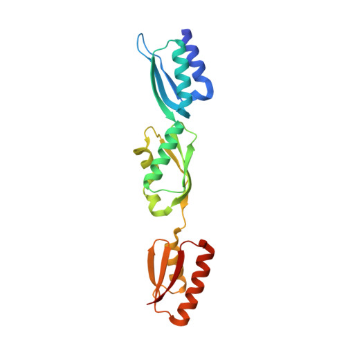
    Injectisomes are multi-protein transmembrane machines allowing pathogenic bacteria to inject effector proteins into eukaryotic host cells, a process called type III secretion. Here we present the first three-dimensional structure of Yersinia enterocolitica and Shigella flexneri injectisomes in situ and the first structural analysis of the Yersinia injectisome. Unexpectedly, basal bodies of injectisomes inside the bacterial cells showed length variations of 20%. The in situ structures of the Y. enterocolitica and S. flexneri injectisomes had similar dimensions and were significantly longer than the isolated structures of related injectisomes. The crystal structure of the inner membrane injectisome component YscD appeared elongated compared to a homologous protein, and molecular dynamics simulations documented its elongation elasticity. The ring-shaped secretin YscC at the outer membrane was stretched by 30-40% in situ, compared to its isolated liposome-embedded conformation. We suggest that elasticity is critical for some two-membrane spanning protein complexes to cope with variations in the intermembrane distance. DOI:http://dx.doi.org/10.7554/eLife.00792.001.


  • Organizational Affiliation
    • Center for Cellular Imaging and NanoAnalytics (C-CINA) , Biozentrum, University of Basel , Basel , Switzerland.

Macromolecule Content 

  • Total Structure Weight: 22.74 kDa 
  • Atom Count: 1,993 
  • Modeled Residue Count: 194 
  • Deposited Residue Count: 198 
  • Unique protein chains: 1

Macromolecules

Find similar proteins by:|  3D Structure
Entity ID: 1
MoleculeChains  Sequence LengthOrganismDetailsImage
YOP PROTEINS TRANSLOCATION PROTEIN D198Yersinia enterocoliticaMutation(s): 1 
UniProt
Find proteins for Q01245 (Yersinia enterocolitica)
Explore Q01245 
Go to UniProtKB:  Q01245
Entity Groups
Sequence Clusters30% Identity50% Identity70% Identity90% Identity95% Identity100% Identity
UniProt GroupQ01245
Sequence Annotations
Expand
Reference Sequence

Experimental Data & Validation

Experimental Data

  • Method: X-RAY DIFFRACTION
  • Resolution: 1.40 Å
  • R-Value Free:  0.226 (Depositor), 0.221 (DCC) 
  • R-Value Work:  0.189 (Depositor), 0.186 (DCC) 
  • R-Value Observed: 0.191 (Depositor) 
Space Group: P 1 21 1
Unit Cell:
Length ( Å )Angle ( ˚ )
a = 37.99α = 90
b = 51.55β = 106.08
c = 50.63γ = 90
Software Package:
Software NamePurpose
PHENIXrefinement
XDSdata reduction
XSCALEdata scaling
Auto-Rickshawphasing

Structure Validation

View Full Validation Report


Entry History 

Deposition Data

Revision History  (Full details and data files)

  • Version 1.0: 2013-04-24
    Type: Initial release
  • Version 1.1: 2013-11-13
    Changes: Database references
  • Version 1.2: 2019-05-08
    Changes: Data collection, Experimental preparation, Other
  • Version 1.3: 2019-05-15
    Changes: Data collection, Experimental preparation
  • Version 1.4: 2024-05-08
    Changes: Data collection, Database references, Derived calculations, Other