4AKT | pdb_00004akt

PatG macrocyclase in complex with peptide


Experimental Data Snapshot

  • Method: X-RAY DIFFRACTION
  • Resolution: 2.63 Å
  • R-Value Free: 
    0.218 (Depositor), 0.221 (DCC) 
  • R-Value Work: 
    0.190 (Depositor), 0.193 (DCC) 
  • R-Value Observed: 
    0.192 (Depositor) 

wwPDB Validation 3D Report Full Report

Validation slider image for 4AKT

This is version 1.3 of the entry. See complete history

Literature

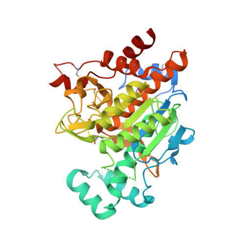
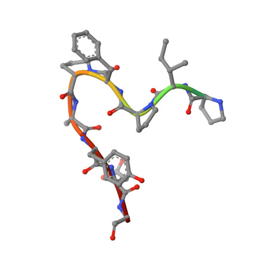
The Mechanism of Patellamide Macrocyclization Revealed by the Characterization of the Patg Macrocyclase Domain.

Koehnke, J.Bent, A.Houssen, W.E.Zollman, D.Morawitz, F.Shirran, S.Vendome, J.Nneoyiegbe, A.F.Trembleau, L.Botting, C.H.Smith, M.C.Jaspars, M.Naismith, J.H.

(2012) Nat Struct Mol Biol 19: 767

  • DOI: https://doi.org/10.1038/nsmb.2340
  • Primary Citation Related Structures: 
    4AKS, 4AKT

  • PubMed Abstract: 

    Peptide macrocycles are found in many biologically active natural products. Their versatility, resistance to proteolysis and ability to traverse membranes has made them desirable molecules. Although technologies exist to synthesize such compounds, the full extent of diversity found among natural macrocycles has yet to be achieved synthetically. Cyanobactins are ribosomal peptide macrocycles encompassing an extraordinarily diverse range of ring sizes, amino acids and chemical modifications. We report the structure, biochemical characterization and initial engineering of the PatG macrocyclase domain of Prochloron sp. from the patellamide pathway that catalyzes the macrocyclization of linear peptides. The enzyme contains insertions in the subtilisin fold to allow it to recognize a three-residue signature, bind substrate in a preorganized and unusual conformation, shield an acyl-enzyme intermediate from water and catalyze peptide bond formation. The ability to macrocyclize a broad range of nonactivated substrates has wide biotechnology applications.


  • Organizational Affiliation
    • Biomedical Sciences Research Complex, University of St Andrews, St Andrews, UK.

Macromolecule Content 

  • Total Structure Weight: 78.22 kDa 
  • Atom Count: 5,108 
  • Modeled Residue Count: 662 
  • Deposited Residue Count: 732 
  • Unique protein chains: 2

Macromolecules

Find similar proteins by:|  3D Structure
Entity ID: 1
MoleculeChains  Sequence LengthOrganismDetailsImage
THIAZOLINE OXIDASE/SUBTILISIN-LIKE PROTEASE
A, B
360Prochloron didemniMutation(s): 1 
UniProt
Find proteins for Q52QJ1 (Prochloron didemni)
Explore Q52QJ1 
Go to UniProtKB:  Q52QJ1
Entity Groups
Sequence Clusters30% Identity50% Identity70% Identity90% Identity95% Identity100% Identity
UniProt GroupQ52QJ1
Sequence Annotations
Expand
Reference Sequence
Find similar proteins by:  Sequence   |   3D Structure  
Entity ID: 2
MoleculeChains  Sequence LengthOrganismDetailsImage
SUBSTRATE ANALOGUE12synthetic constructMutation(s): 0 

Experimental Data & Validation

Experimental Data

  • Method: X-RAY DIFFRACTION
  • Resolution: 2.63 Å
  • R-Value Free:  0.218 (Depositor), 0.221 (DCC) 
  • R-Value Work:  0.190 (Depositor), 0.193 (DCC) 
  • R-Value Observed: 0.192 (Depositor) 
Space Group: C 1 2 1
Unit Cell:
Length ( Å )Angle ( ˚ )
a = 135.63α = 90
b = 67.32β = 116.76
c = 137.87γ = 90
Software Package:
Software NamePurpose
REFMACrefinement
PHASERphasing

Structure Validation

View Full Validation Report


Entry History 

Revision History  (Full details and data files)

  • Version 1.0: 2012-07-18
    Type: Initial release
  • Version 1.1: 2012-08-22
    Changes: Database references
  • Version 1.2: 2013-11-06
    Changes: Structure summary
  • Version 1.3: 2024-11-20
    Changes: Data collection, Database references, Other, Structure summary