4AKK

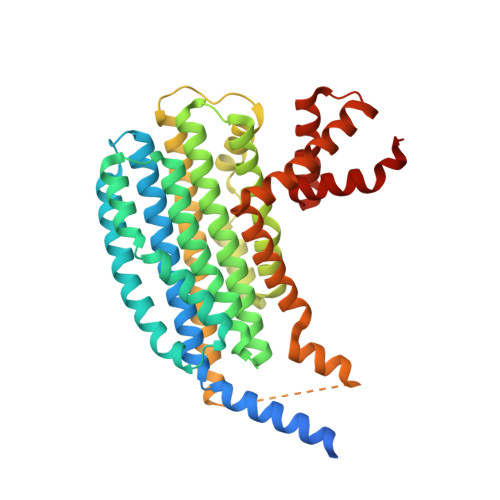
Structure of the NasR transcription antiterminator


Experimental Data Snapshot

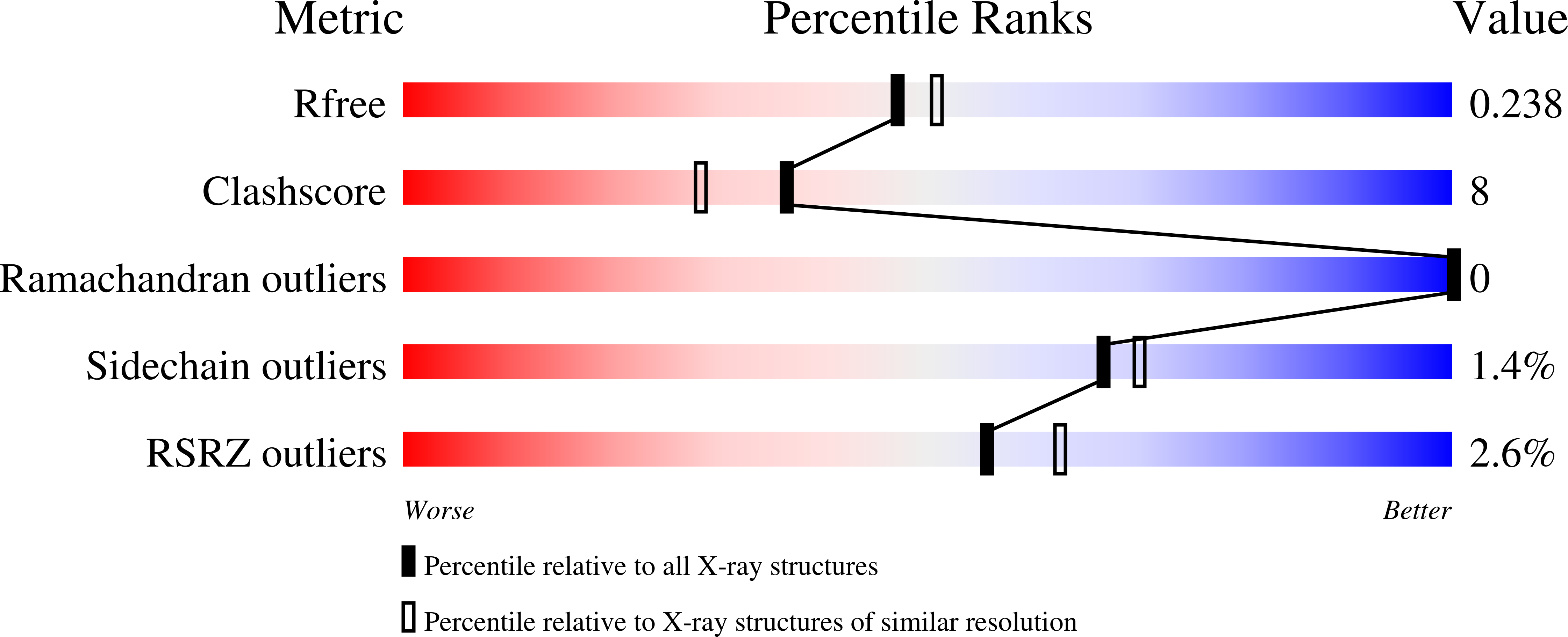
  • Method: X-RAY DIFFRACTION
  • Resolution: 2.15 Å
  • R-Value Free: 0.241 
  • R-Value Work: 0.200 
  • R-Value Observed: 0.203 

wwPDB Validation   3D Report Full Report


This is version 1.4 of the entry. See complete history


Literature

The Structure of the Nasr Transcription Antiterminator Reveals a One-Component System with a Nit Nitrate Receptor Coupled to an Antar RNA-Binding Effector.

Boudes, M.Lazar, N.Graille, M.Durand, D.Gaidenko, T.A.Stewart, V.Van Tilbeurgh, H.

(2012) Mol Microbiol 85: 431

  • DOI: https://doi.org/10.1111/j.1365-2958.2012.08111.x
  • Primary Citation of Related Structures:  
    4AKK

  • PubMed Abstract: 

    The nitrate- and nitrite-sensing NIT domain is present in diverse signal-transduction proteins across a wide range of bacterial species. NIT domain function was established through analysis of the Klebsiella oxytoca NasR protein, which controls expression of the nasF operon encoding enzymes for nitrite and nitrate assimilation. In the presence of nitrate or nitrite, the NasR protein inhibits transcription termination at the factor-independent terminator site in the nasF operon transcribed leader region. We present here the crystal structure of the intact NasR protein in the apo state. The dimeric all-helical protein contains a large amino-terminal NIT domain that associates two four-helix bundles, and a carboxyl-terminal ANTAR (AmiR and NasR transcription antitermination regulator) domain. The analysis reveals unexpectedly that the NIT domain is structurally similar to the periplasmic input domain of the NarX two-component sensor that regulates nitrate and nitrite respiration. This similarity suggests that the NIT domain binds nitrate and nitrite between two invariant arginyl residues located on adjacent alpha helices, and results from site-specific mutagenesis showed that these residues are critical for NasR function. The resulting structural movements in the NIT domain would provoke an active configuration of the ANTAR domains necessary for specific leader mRNA binding.


  • Organizational Affiliation

    IBBMC-CNRS UMR8619, Bât. 430, Université Paris-Sud, 91405 Orsay, France.


Macromolecules
Find similar proteins by:  (by identity cutoff)  |  3D Structure
Entity ID: 1
MoleculeChains Sequence LengthOrganismDetailsImage
NITRATE REGULATORY PROTEIN
A, B
423Klebsiella oxytocaMutation(s): 0 
UniProt
Find proteins for Q48468 (Klebsiella oxytoca)
Explore Q48468 
Go to UniProtKB:  Q48468
Entity Groups  
Sequence Clusters30% Identity50% Identity70% Identity90% Identity95% Identity100% Identity
UniProt GroupQ48468
Sequence Annotations
Expand
  • Reference Sequence
Small Molecules
Ligands 1 Unique
IDChains Name / Formula / InChI Key2D Diagram3D Interactions
EDO
Query on EDO

Download Ideal Coordinates CCD File 
C [auth A]
D [auth A]
E [auth A]
F [auth B]
G [auth B]
C [auth A],
D [auth A],
E [auth A],
F [auth B],
G [auth B],
H [auth B],
I [auth B],
J [auth B],
K [auth B]
1,2-ETHANEDIOL
C2 H6 O2
LYCAIKOWRPUZTN-UHFFFAOYSA-N
Experimental Data & Validation

Experimental Data

  • Method: X-RAY DIFFRACTION
  • Resolution: 2.15 Å
  • R-Value Free: 0.241 
  • R-Value Work: 0.200 
  • R-Value Observed: 0.203 
  • Space Group: C 1 2 1
Unit Cell:
Length ( Å )Angle ( ˚ )
a = 159.23α = 90
b = 113.1β = 109.3
c = 59.35γ = 90
Software Package:
Software NamePurpose
PHENIXrefinement
XDSdata reduction
XSCALEdata scaling
SHARPphasing

Structure Validation

View Full Validation Report


Entry History 

Deposition Data

Revision History  (Full details and data files)

  • Version 1.0: 2012-06-20
    Type: Initial release
  • Version 1.1: 2012-08-08
    Changes: Database references
  • Version 1.2: 2019-01-30
    Changes: Data collection, Experimental preparation, Other
  • Version 1.3: 2019-02-06
    Changes: Data collection, Experimental preparation
  • Version 1.4: 2024-05-08
    Changes: Data collection, Database references, Derived calculations, Other