4AKE | pdb_00004ake

ADENYLATE KINASE


Experimental Data Snapshot

  • Method: X-RAY DIFFRACTION
  • Resolution: 2.20 Å
  • R-Value Work: 
    0.183 (Depositor), 0.178 (DCC) 
  • R-Value Observed: 
    0.183 (Depositor) 

wwPDB Validation 3D Report Full Report

Validation slider image for 4AKE

This is version 1.3 of the entry. See complete history

Literature

Adenylate kinase motions during catalysis: an energetic counterweight balancing substrate binding.

Muller, C.W.Schlauderer, G.J.Reinstein, J.Schulz, G.E.

(1996) Structure 4: 147-156

  • DOI: https://doi.org/10.1016/s0969-2126(96)00018-4
  • Primary Citation Related Structures: 
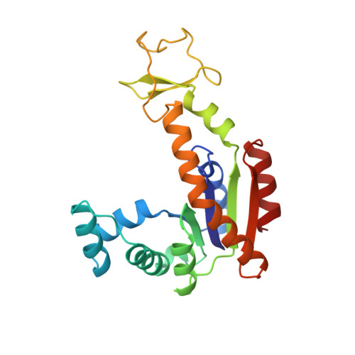
    4AKE

  • PubMed Abstract: 

    Adenylate kinases undergo large conformational changes during their catalytic cycle. Because these changes have been studied by comparison of structures from different species, which share approximately one-third of their residues, only rough descriptions have been possible to date. We have solved the structure of unligated adenylate kinase from Escherichia coli at 2.2 degree resolution and compared it with the high-resolution structure of the same enzyme ligated with an inhibitor mimicking both substrates, ATP and AMP. This comparison shows that, upon substrate binding, the enzyme increases its chain mobility in a region remote from the active center. As this region 'solidifies' again on substrate release, we propose that it serves as a 'counterweight' balancing the substrate binding energy. The comparison of two very different conformations of the same polypeptide chain revealed kinematic details of the catalytic cycle. Moreover, it indicated that there exists an energetic counterweight compensating the substrate binding energy required for specificity. This counterweight prevents the enzyme from dropping into a rate-reducing energy well along the reaction coordinate.


  • Organizational Affiliation
    • Institut für Organische Chemie und Biochemie, 79104-Freiburg im Breisgau, Germany.

Macromolecule Content 

  • Total Structure Weight: 47.24 kDa 
  • Atom Count: 3,459 
  • Modeled Residue Count: 428 
  • Deposited Residue Count: 428 
  • Unique protein chains: 1

Macromolecules

Find similar proteins by:|  3D Structure
Entity ID: 1
MoleculeChains  Sequence LengthOrganismDetailsImage
ADENYLATE KINASE
A, B
214Escherichia coliMutation(s): 0 
EC: 2.7.4.3
UniProt
Find proteins for P69441 (Escherichia coli (strain K12))
Explore P69441 
Go to UniProtKB:  P69441
Entity Groups
Sequence Clusters30% Identity50% Identity70% Identity90% Identity95% Identity100% Identity
UniProt GroupP69441
Sequence Annotations
Expand
Reference Sequence

Experimental Data & Validation

Experimental Data

  • Method: X-RAY DIFFRACTION
  • Resolution: 2.20 Å
  • R-Value Work:  0.183 (Depositor), 0.178 (DCC) 
  • R-Value Observed: 0.183 (Depositor) 
Space Group: P 1
Unit Cell:
Length ( Å )Angle ( ˚ )
a = 31.8α = 67.7
b = 54.5β = 77.9
c = 71.3γ = 88.4
Software Package:
Software NamePurpose
X-PLORmodel building
X-PLORrefinement
XDSdata reduction
X-PLORphasing

Structure Validation

View Full Validation Report


Entry History 

Deposition Data

Revision History  (Full details and data files)

  • Version 1.0: 1996-06-10
    Type: Initial release
  • Version 1.1: 2008-03-25
    Changes: Version format compliance
  • Version 1.2: 2011-07-13
    Changes: Version format compliance
  • Version 1.3: 2024-02-28
    Changes: Data collection, Database references, Other