4AD0 | pdb_00004ad0

Structure of the GH99 endo-alpha-mannosidase from Bacteriodes thetaiotaomicron in complex with BIS-TRIS-Propane


Experimental Data Snapshot

  • Method: X-RAY DIFFRACTION
  • Resolution: 2.09 Å
  • R-Value Free: 
    0.237 (Depositor), 0.203 (DCC) 
  • R-Value Work: 
    0.181 (Depositor), 0.158 (DCC) 
  • R-Value Observed: 
    0.184 (Depositor) 

Starting Model: experimental
View more details

wwPDB Validation 3D Report Full Report

Validation slider image for 4AD0

This is version 1.2 of the entry. See complete history

Literature

Structural and Mechanistic Insight Into N-Glycan Processing by Endo-Alpha-Mannosidase.

Thompson, A.J.Williams, R.J.Hakki, Z.Alonzi, D.S.Wennekes, T.Gloster, T.M.Songsrirote, K.Thomas-Oates, J.E.Wrodnigg, T.M.Spreitz, J.Stutz, A.E.Butters, T.D.Williams, S.J.Davies, G.J.

(2012) Proc Natl Acad Sci U S A 109: 781

  • DOI: https://doi.org/10.1073/pnas.1111482109
  • Primary Citation Related Structures: 
    4ACY, 4ACZ, 4AD0, 4AD1, 4AD2, 4AD3, 4AD4, 4AD5

  • PubMed Abstract: 

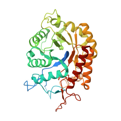
    N-linked glycans play key roles in protein folding, stability, and function. Biosynthetic modification of N-linked glycans, within the endoplasmic reticulum, features sequential trimming and readornment steps. One unusual enzyme, endo-α-mannosidase, cleaves mannoside linkages internally within an N-linked glycan chain, short circuiting the classical N-glycan biosynthetic pathway. Here, using two bacterial orthologs, we present the first structural and mechanistic dissection of endo-α-mannosidase. Structures solved at resolutions 1.7-2.1 Å reveal a (β/α)(8) barrel fold in which the catalytic center is present in a long substrate-binding groove, consistent with cleavage within the N-glycan chain. Enzymatic cleavage of authentic Glc(1/3)Man(9)GlcNAc(2) yields Glc(1/3)-Man. Using the bespoke substrate α-Glc-1,3-α-Man fluoride, the enzyme was shown to act with retention of anomeric configuration. Complexes with the established endo-α-mannosidase inhibitor α-Glc-1,3-deoxymannonojirimycin and a newly developed inhibitor, α-Glc-1,3-isofagomine, and with the reducing-end product α-1,2-mannobiose structurally define the -2 to +2 subsites of the enzyme. These structural and mechanistic data provide a foundation upon which to develop new enzyme inhibitors targeting the hijacking of N-glycan synthesis in viral disease and cancer.


  • Organizational Affiliation
    • Department of Chemistry, University of York, Heslington, York YO10 5DD, United Kingdom.

Macromolecule Content 

  • Total Structure Weight: 174.55 kDa 
  • Atom Count: 10,639 
  • Modeled Residue Count: 1,285 
  • Deposited Residue Count: 1,528 
  • Unique protein chains: 1

Macromolecules

Find similar proteins by:|  3D Structure
Entity ID: 1
MoleculeChains  Sequence LengthOrganismDetailsImage
ENDO-ALPHA-MANNOSIDASE
A, B, C, D
382Bacteroides thetaiotaomicron VPI-5482Mutation(s): 0 
EC: 3.2.1.130
UniProt
Find proteins for Q8A109 (Bacteroides thetaiotaomicron (strain ATCC 29148 / DSM 2079 / JCM 5827 / CCUG 10774 / NCTC 10582 / VPI-5482 / E50))
Explore Q8A109 
Go to UniProtKB:  Q8A109
Entity Groups
Sequence Clusters30% Identity50% Identity70% Identity90% Identity95% Identity100% Identity
UniProt GroupQ8A109
Sequence Annotations
Expand
Reference Sequence

Small Molecules

Ligands 2 Unique
IDChains Name / Formula / InChI Key2D Diagram3D Interactions
B3P

Query on B3P


Download:Ideal Coordinates CCD File
E [auth A],
G [auth B],
I [auth C],
K [auth D]
2-[3-(2-HYDROXY-1,1-DIHYDROXYMETHYL-ETHYLAMINO)-PROPYLAMINO]-2-HYDROXYMETHYL-PROPANE-1,3-DIOL
C11 H26 N2 O6
HHKZCCWKTZRCCL-UHFFFAOYSA-N
GOL

Query on GOL


Download:Ideal Coordinates CCD File
F [auth A],
H [auth B],
J [auth C],
L [auth D]
GLYCEROL
C3 H8 O3
PEDCQBHIVMGVHV-UHFFFAOYSA-N

Experimental Data & Validation

Experimental Data

  • Method: X-RAY DIFFRACTION
  • Resolution: 2.09 Å
  • R-Value Free:  0.237 (Depositor), 0.203 (DCC) 
  • R-Value Work:  0.181 (Depositor), 0.158 (DCC) 
  • R-Value Observed: 0.184 (Depositor) 
Space Group: P 1 21 1
Unit Cell:
Length ( Å )Angle ( ˚ )
a = 65.993α = 90
b = 168.428β = 117.08
c = 72.433γ = 90
Software Package:
Software NamePurpose
REFMACrefinement
HKL-2000data reduction
SCALEPACKdata scaling
MOLREPphasing

Structure Validation

View Full Validation Report


Entry History 

Revision History  (Full details and data files)

  • Version 1.0: 2012-02-01
    Type: Initial release
  • Version 1.1: 2012-02-29
    Changes: Other
  • Version 1.2: 2023-12-20
    Changes: Data collection, Database references, Derived calculations, Other, Refinement description