4A9V | pdb_00004a9v

Pseudomonas fluorescens PhoX


Experimental Data Snapshot

  • Method: X-RAY DIFFRACTION
  • Resolution: 1.10 Å
  • R-Value Free: 
    0.117 (Depositor), 0.146 (DCC) 
  • R-Value Work: 
    0.137 (Depositor), 0.132 (DCC) 
  • R-Value Observed: 
    0.118 (Depositor) 

Starting Model: experimental
View more details

wwPDB Validation 3D Report Full Report

Validation slider image for 4A9V

This is version 1.4 of the entry. See complete history

Literature

A Complex Iron-Calcium Cofactor Catalyzing Phosphotransfer Chemistry

Yong, S.C.Roversi, P.Lillington, J.Rodriguez, F.Krehenbrink, M.Zeldin, O.B.Garman, E.F.Lea, S.M.Berks, B.C.

(2014) Science 345: 1170

  • DOI: https://doi.org/10.1126/science.1254237
  • Primary Citation Related Structures: 
    3ZWU, 4A9V, 4A9X, 4ALF, 4AMF

  • PubMed Abstract: 

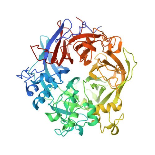
    Alkaline phosphatases play a crucial role in phosphate acquisition by microorganisms. To expand our understanding of catalysis by this class of enzymes, we have determined the structure of the widely occurring microbial alkaline phosphatase PhoX. The enzyme contains a complex active-site cofactor comprising two antiferromagnetically coupled ferric iron ions (Fe(3+)), three calcium ions (Ca(2+)), and an oxo group bridging three of the metal ions. Notably, the main part of the cofactor resembles synthetic oxide-centered triangular metal complexes. Structures of PhoX-ligand complexes reveal how the active-site metal ions bind substrate and implicate the cofactor oxo group in the catalytic mechanism. The presence of iron in PhoX raises the possibility that iron bioavailability limits microbial phosphate acquisition.


  • Organizational Affiliation
    • Department of Biochemistry, University of Oxford, South Parks Road, Oxford OX1 3QU, United Kingdom.

Macromolecule Content 

  • Total Structure Weight: 65.5 kDa 
  • Atom Count: 5,585 
  • Modeled Residue Count: 581 
  • Deposited Residue Count: 592 
  • Unique protein chains: 1

Macromolecules

Find similar proteins by:|  3D Structure
Entity ID: 1
MoleculeChains  Sequence LengthOrganismDetailsImage
PHOX592Pseudomonas fluorescens Pf0-1Mutation(s): 0 
EC: 3.1.3.1
UniProt
Find proteins for Q3K5N8 (Pseudomonas fluorescens (strain Pf0-1))
Explore Q3K5N8 
Go to UniProtKB:  Q3K5N8
Entity Groups
Sequence Clusters30% Identity50% Identity70% Identity90% Identity95% Identity100% Identity
UniProt GroupQ3K5N8
Sequence Annotations
Expand
Reference Sequence

Small Molecules

Ligands 6 Unique
IDChains Name / Formula / InChI Key2D Diagram3D Interactions
FEO

Query on FEO


Download:Ideal Coordinates CCD File
S [auth A]MU-OXO-DIIRON
Fe2 O
NPMYUMBHPJGBFA-UHFFFAOYSA-N
EDO

Query on EDO


Download:Ideal Coordinates CCD File
H [auth A]
I [auth A]
J [auth A]
K [auth A]
L [auth A]
H [auth A],
I [auth A],
J [auth A],
K [auth A],
L [auth A],
M [auth A],
N [auth A],
O [auth A],
P [auth A]
1,2-ETHANEDIOL
C2 H6 O2
LYCAIKOWRPUZTN-UHFFFAOYSA-N
CA

Query on CA


Download:Ideal Coordinates CCD File
Q [auth A],
R [auth A]
CALCIUM ION
Ca
BHPQYMZQTOCNFJ-UHFFFAOYSA-N
CL

Query on CL


Download:Ideal Coordinates CCD File
B [auth A]CHLORIDE ION
Cl
VEXZGXHMUGYJMC-UHFFFAOYSA-M
NA

Query on NA


Download:Ideal Coordinates CCD File
E [auth A],
F [auth A]
SODIUM ION
Na
FKNQFGJONOIPTF-UHFFFAOYSA-N
LI

Query on LI


Download:Ideal Coordinates CCD File
C [auth A],
D [auth A],
G [auth A]
LITHIUM ION
Li
HBBGRARXTFLTSG-UHFFFAOYSA-N

Experimental Data & Validation

Experimental Data

  • Method: X-RAY DIFFRACTION
  • Resolution: 1.10 Å
  • R-Value Free:  0.117 (Depositor), 0.146 (DCC) 
  • R-Value Work:  0.137 (Depositor), 0.132 (DCC) 
  • R-Value Observed: 0.118 (Depositor) 
Space Group: P 21 21 21
Unit Cell:
Length ( Å )Angle ( ˚ )
a = 70.82α = 90
b = 75.98β = 90
c = 114.97γ = 90
Software Package:
Software NamePurpose
REFMACrefinement
XDSdata reduction
SCALAdata scaling
PHASERphasing

Structure Validation

View Full Validation Report


Entry History 

Deposition Data

Revision History  (Full details and data files)

  • Version 1.0: 2012-12-05
    Type: Initial release
  • Version 1.1: 2014-09-10
    Changes: Database references
  • Version 1.2: 2014-09-17
    Changes: Database references
  • Version 1.3: 2018-01-24
    Changes: Source and taxonomy
  • Version 1.4: 2023-12-20
    Changes: Data collection, Database references, Derived calculations, Other, Refinement description