4A95 | pdb_00004a95

Plasmodium vivax N-myristoyltransferase with quinoline inhibitor


Experimental Data Snapshot

  • Method: X-RAY DIFFRACTION
  • Resolution: 1.55 Å
  • R-Value Free: 
    0.236 (Depositor), 0.235 (DCC) 
  • R-Value Work: 
    0.186 (Depositor), 0.186 (DCC) 
  • R-Value Observed: 
    0.189 (Depositor) 

Starting Model: other
View more details

wwPDB Validation 3D Report Full Report

Validation slider image for 4A95

Ligand Structure Quality Assessment 


This is version 1.2 of the entry. See complete history

Literature

Discovery of Plasmodium Vivax N-Myristoyltransferase Inhibitors: Screening, Synthesis, and Structural Characterization of Their Binding Mode.

Goncalves, V.Brannigan, J.A.Whalley, D.Ansell, K.H.Saxty, B.Holder, A.A.Wilkinson, A.J.Tate, E.W.Leatherbarrow, R.J.

(2012) J Med Chem 55: 3578

  • DOI: https://doi.org/10.1021/jm300040p
  • Primary Citation Related Structures: 
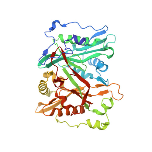
    4A95

  • PubMed Abstract: 

    N-Myristoyltransferase (NMT) is a prospective drug target against parasitic protozoa. Herein we report the successful discovery of a series of Plasmodium vivax NMT inhibitors by high-throughput screening. A high-resolution crystal structure of the hit compound in complex with NMT was obtained, allowing understanding of its novel binding mode. A set of analogues was designed and tested to define the chemical groups relevant for activity and selectivity.


  • Organizational Affiliation
    • Department of Chemistry, Imperial College London, London SW7 2AZ, United Kingdom.

Macromolecule Content 

  • Total Structure Weight: 139.03 kDa 
  • Atom Count: 11,498 
  • Modeled Residue Count: 1,134 
  • Deposited Residue Count: 1,152 
  • Unique protein chains: 1

Macromolecules

Find similar proteins by:|  3D Structure
Entity ID: 1
MoleculeChains  Sequence LengthOrganismDetailsImage
GLYCYLPEPTIDE N-TETRADECANOYLTRANSFERASE
A, B, C
384Plasmodium vivaxMutation(s): 0 
EC: 2.3.1.97
UniProt
Find proteins for A5K1A2 (Plasmodium vivax (strain Salvador I))
Explore A5K1A2 
Go to UniProtKB:  A5K1A2
Entity Groups
Sequence Clusters30% Identity50% Identity70% Identity90% Identity95% Identity100% Identity
UniProt GroupA5K1A2
Sequence Annotations
Expand
Reference Sequence

Small Molecules

Ligands 5 Unique
IDChains Name / Formula / InChI Key2D Diagram3D Interactions
NHW

Query on NHW


Download:Ideal Coordinates CCD File
E [auth A],
I [auth B],
N [auth C]
2-oxopentadecyl-CoA
C36 H64 N7 O17 P3 S
JKWHUJMJVNMKEF-UOCZADIYSA-J
9MT

Query on 9MT


Download:Ideal Coordinates CCD File
D [auth A],
H [auth B],
M [auth C]
3-(3-BUTYL-6-METHOXY-2-METHYL-QUINOLIN-4-YL)SULFANYLPROPANENITRILE
C18 H22 N2 O S
UJFIVLIFWNFOIZ-UHFFFAOYSA-N
SO4

Query on SO4


Download:Ideal Coordinates CCD File
J [auth B]SULFATE ION
O4 S
QAOWNCQODCNURD-UHFFFAOYSA-L
CL

Query on CL


Download:Ideal Coordinates CCD File
F [auth A],
K [auth B],
O [auth C]
CHLORIDE ION
Cl
VEXZGXHMUGYJMC-UHFFFAOYSA-M
MG

Query on MG


Download:Ideal Coordinates CCD File
G [auth A],
L [auth B],
P [auth C]
MAGNESIUM ION
Mg
JLVVSXFLKOJNIY-UHFFFAOYSA-N

Experimental Data & Validation

Experimental Data

  • Method: X-RAY DIFFRACTION
  • Resolution: 1.55 Å
  • R-Value Free:  0.236 (Depositor), 0.235 (DCC) 
  • R-Value Work:  0.186 (Depositor), 0.186 (DCC) 
  • R-Value Observed: 0.189 (Depositor) 
Space Group: P 21 21 21
Unit Cell:
Length ( Å )Angle ( ˚ )
a = 57.49α = 90
b = 121.56β = 90
c = 179.01γ = 90
Software Package:
Software NamePurpose
REFMACrefinement
XDSdata reduction
XSCALEdata scaling
MOLREPphasing

Structure Validation

View Full Validation Report


Ligand Structure Quality Assessment 


Entry History 

Deposition Data

Revision History  (Full details and data files)

  • Version 1.0: 2012-06-27
    Type: Initial release
  • Version 1.1: 2012-07-18
    Changes: Atomic model, Derived calculations, Non-polymer description, Refinement description
  • Version 1.2: 2024-05-01
    Changes: Data collection, Database references, Derived calculations, Other, Refinement description