4A6G | pdb_00004a6g

N-acyl amino acid racemase from Amycalotopsis sp. Ts-1-60: G291D- F323Y mutant in complex with N-acetyl methionine


Experimental Data Snapshot

  • Method: X-RAY DIFFRACTION
  • Resolution: 2.71 Å
  • R-Value Free: 
    0.210 (Depositor), 0.207 (DCC) 
  • R-Value Work: 
    0.155 (Depositor), 0.153 (DCC) 
  • R-Value Observed: 
    0.158 (Depositor) 

Starting Model: experimental
View more details

wwPDB Validation 3D Report Full Report

Validation slider image for 4A6G

Ligand Structure Quality Assessment 


This is version 1.3 of the entry. See complete history

Literature

An Improved Racemase/Acylase Biotransformation for the Preparation of Enantiomerically Pure Amino Acids.

Baxter, S.Royer, S.Grogan, G.Brown, F.Holt-Tiffin, K.E.Taylor, I.N.Fotheringham, I.G.Campopiano, D.J.

(2012) J Am Chem Soc 134: 19310

  • DOI: https://doi.org/10.1021/ja305438y
  • Primary Citation Related Structures: 
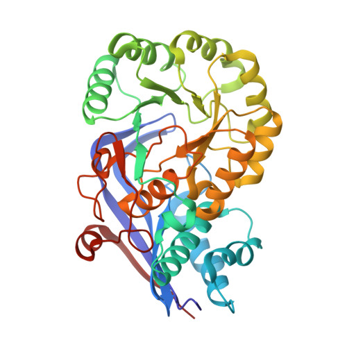
    4A6G

  • PubMed Abstract: 

    Using directed evolution, a variant N-acetyl amino acid racemase (NAAAR G291D/F323Y) has been developed with up to 6-fold higher activity than the wild-type on a range of N-acetylated amino acids. The variant has been coupled with an enantiospecific acylase to give a preparative scale dynamic kinetic resolution which allows 98% conversion of N-acetyl-DL-allylglycine into D-allylglycine in 18 h at high substrate concentrations (50 g L(-1)). This is the first example of NAAAR operating under conditions which would allow it to be successfully used on an industrial scale for the production of enantiomerically pure α-amino acids. X-ray crystal analysis of the improved NAAAR variant allowed a comparison with the wild-type enzyme. We postulate that a network of novel interactions that result from the introduction of the two side chains is the source of improved catalytic performance.


  • Organizational Affiliation
    • The EastChem School of Chemistry, Joseph Black Building, The University of Edinburgh, Edinburgh, EH9 3JJ, UK.

Macromolecule Content 

  • Total Structure Weight: 158.95 kDa 
  • Atom Count: 11,214 
  • Modeled Residue Count: 1,469 
  • Deposited Residue Count: 1,472 
  • Unique protein chains: 1

Macromolecules

Find similar proteins by:|  3D Structure
Entity ID: 1
MoleculeChains  Sequence LengthOrganismDetailsImage
N-ACYLAMINO ACID RACEMASE
A, B, C, D
368Amycolatopsis sp.Mutation(s): 2 
EC: 4.2.1.113 (PDB Primary Data), 5.1.1 (UniProt)
UniProt
Find proteins for Q44244 (Amycolatopsis sp)
Explore Q44244 
Go to UniProtKB:  Q44244
Entity Groups
Sequence Clusters30% Identity50% Identity70% Identity90% Identity95% Identity100% Identity
UniProt GroupQ44244
Sequence Annotations
Expand
Reference Sequence

Small Molecules

Ligands 2 Unique
IDChains Name / Formula / InChI Key2D Diagram3D Interactions
AME

Query on AME


Download:Ideal Coordinates CCD File
E [auth A],
G [auth B],
I [auth C],
K [auth D]
N-ACETYLMETHIONINE
C7 H13 N O3 S
XUYPXLNMDZIRQH-LURJTMIESA-N
MG

Query on MG


Download:Ideal Coordinates CCD File
F [auth A],
H [auth B],
J [auth C],
L [auth D]
MAGNESIUM ION
Mg
JLVVSXFLKOJNIY-UHFFFAOYSA-N

Experimental Data & Validation

Experimental Data

  • Method: X-RAY DIFFRACTION
  • Resolution: 2.71 Å
  • R-Value Free:  0.210 (Depositor), 0.207 (DCC) 
  • R-Value Work:  0.155 (Depositor), 0.153 (DCC) 
  • R-Value Observed: 0.158 (Depositor) 
Space Group: H 3 2
Unit Cell:
Length ( Å )Angle ( ˚ )
a = 216.71α = 90
b = 216.71β = 90
c = 261.1γ = 120
Software Package:
Software NamePurpose
REFMACrefinement
xia2data reduction
xia2data scaling
PHASERphasing

Structure Validation

View Full Validation Report


Ligand Structure Quality Assessment 


Entry History 

Deposition Data

Revision History  (Full details and data files)

  • Version 1.0: 2012-11-14
    Type: Initial release
  • Version 1.1: 2012-11-21
    Changes: Database references
  • Version 1.2: 2012-12-19
    Changes: Database references
  • Version 1.3: 2023-12-20
    Changes: Data collection, Database references, Derived calculations, Other, Refinement description