4A2C | pdb_00004a2c

Crystal structure of galactitol-1-phosphate dehydrogenase from Escherichia coli


Experimental Data Snapshot

  • Method: X-RAY DIFFRACTION
  • Resolution: 1.87 Å
  • R-Value Free: 
    0.273 (Depositor), 0.269 (DCC) 
  • R-Value Work: 
    0.213 (Depositor), 0.208 (DCC) 
  • R-Value Observed: 
    0.216 (Depositor) 

Starting Model: experimental
View more details

wwPDB Validation 3D Report Full Report

Validation slider image for 4A2C

This is version 1.3 of the entry. See complete history

Literature

The Crystal Structure of Galactitol-1-Phosphate 5-Dehydrogenase from Escherichia Coli K12 Provides Insights Into its Anomalous Behavior on Imac Processes

Esteban-Torres, M.Alvarez, Y.Acebron, I.De Las Rivas, B.Munoz, R.Kohring, G.-W.Roa, A.M.Sobrino, M.Mancheno, J.M.

(2012) FEBS Lett 586: 3127

  • DOI: https://doi.org/10.1016/j.febslet.2012.07.073
  • Primary Citation Related Structures: 
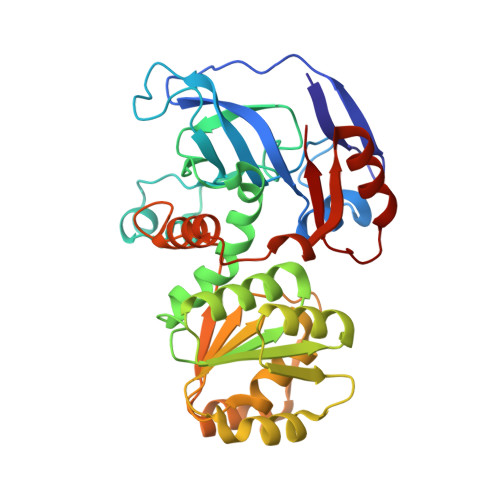
    4A2C

  • PubMed Abstract: 

    Endogenous galactitol-1-phosphate 5-dehydrogenase (GPDH) (EC 1.1.1.251) from Escherichia coli spontaneously interacts with Ni(2+)-NTA matrices becoming a potential contaminant for recombinant, target His-tagged proteins. Purified recombinant, untagged GPDH (rGPDH) converted galactitol into tagatose, and d-tagatose-6-phosphate into galactitol-1-phosphate, in a Zn(2+)- and NAD(H)-dependent manner and readily crystallized what has permitted to solve its crystal structure. In contrast, N-terminally His-tagged GPDH was marginally stable and readily aggregated. The structure of rGPDH revealed metal-binding sites characteristic from the medium-chain dehydrogenase/reductase protein superfamily which may explain its ability to interact with immobilized metals. The structure also provides clues on the harmful effects of the N-terminal His-tag.


  • Organizational Affiliation
    • Laboratorio de Biotecnología Bacteriana, Instituto Ciencia y Tecnología de Alimentos y Nutrición (ICTAN), CSIC, Madrid, Spain.

Macromolecule Content 

  • Total Structure Weight: 75.17 kDa 
  • Atom Count: 5,923 
  • Modeled Residue Count: 692 
  • Deposited Residue Count: 692 
  • Unique protein chains: 1

Macromolecules

Find similar proteins by:|  3D Structure
Entity ID: 1
MoleculeChains  Sequence LengthOrganismDetailsImage
GALACTITOL-1-PHOSPHATE 5-DEHYDROGENASE
A, B
346Escherichia coli K-12Mutation(s): 0 
EC: 1.1.1.251
UniProt
Find proteins for P0A9S3 (Escherichia coli (strain K12))
Explore P0A9S3 
Go to UniProtKB:  P0A9S3
Entity Groups
Sequence Clusters30% Identity50% Identity70% Identity90% Identity95% Identity100% Identity
UniProt GroupP0A9S3
Sequence Annotations
Expand
Reference Sequence

Experimental Data & Validation

Experimental Data

  • Method: X-RAY DIFFRACTION
  • Resolution: 1.87 Å
  • R-Value Free:  0.273 (Depositor), 0.269 (DCC) 
  • R-Value Work:  0.213 (Depositor), 0.208 (DCC) 
  • R-Value Observed: 0.216 (Depositor) 
Space Group: P 1 21 1
Unit Cell:
Length ( Å )Angle ( ˚ )
a = 43.72α = 90
b = 77.14β = 95.36
c = 107.51γ = 90
Software Package:
Software NamePurpose
PHENIXrefinement
MOSFLMdata reduction
SCALAdata scaling
PHASERphasing

Structure Validation

View Full Validation Report


Entry History 

Deposition Data

Revision History  (Full details and data files)

  • Version 1.0: 2012-08-08
    Type: Initial release
  • Version 1.1: 2012-08-22
    Changes: Database references
  • Version 1.2: 2012-10-03
    Changes: Database references
  • Version 1.3: 2023-12-20
    Changes: Data collection, Database references, Derived calculations, Other, Refinement description