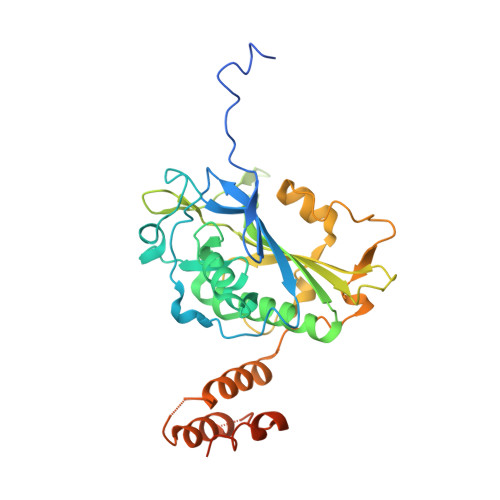
4A1N | pdb_00004a1n

Human Mitochondrial endo-exonuclease


Experimental Data Snapshot

  • Method: X-RAY DIFFRACTION
  • Resolution: 2.80 Å
  • R-Value Free: 
    0.225 (Depositor), 0.207 (DCC) 
  • R-Value Work: 
    0.175 (Depositor) 
  • R-Value Observed: 
    0.178 (Depositor) 

Starting Model: experimental
View more details

wwPDB Validation 3D Report Full Report

Validation slider image for 4A1N

This is version 1.1 of the entry. See complete history

Macromolecule Content 

  • Total Structure Weight: 38.1 kDa 
  • Atom Count: 2,379 
  • Modeled Residue Count: 290 
  • Deposited Residue Count: 335 
  • Unique protein chains: 1

Macromolecules

Find similar proteins by:|  3D Structure
Entity ID: 1
MoleculeChains  Sequence LengthOrganismDetailsImage
NUCLEASE EXOG, MITOCHONDRIAL335Homo sapiensMutation(s): 0 
EC: 3.1.30
UniProt & NIH Common Fund Data Resources
Find proteins for Q9Y2C4 (Homo sapiens)
Explore Q9Y2C4 
Go to UniProtKB:  Q9Y2C4
PHAROS:  Q9Y2C4
GTEx:  ENSG00000157036 
Entity Groups
Sequence Clusters30% Identity50% Identity70% Identity90% Identity95% Identity100% Identity
UniProt GroupQ9Y2C4
Sequence Annotations
Expand
Reference Sequence

Experimental Data & Validation

Experimental Data

  • Method: X-RAY DIFFRACTION
  • Resolution: 2.80 Å
  • R-Value Free:  0.225 (Depositor), 0.207 (DCC) 
  • R-Value Work:  0.175 (Depositor) 
  • R-Value Observed: 0.178 (Depositor) 
Space Group: P 31 2 1
Unit Cell:
Length ( Å )Angle ( ˚ )
a = 103.18α = 90
b = 103.18β = 90
c = 83.89γ = 120
Software Package:
Software NamePurpose
BUSTERrefinement
XDSdata reduction
XSCALEdata scaling
PHASERphasing

Structure Validation

View Full Validation Report


Entry History 

Revision History  (Full details and data files)

  • Version 1.0: 2012-02-29
    Type: Initial release
  • Version 1.1: 2023-12-20
    Changes: Data collection, Database references, Derived calculations, Other, Refinement description