43EG | pdb_000043eg

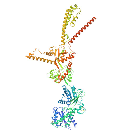
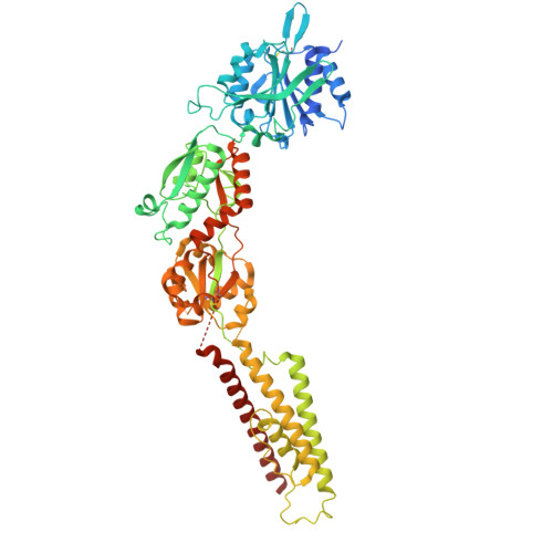
Cryo-EM structure of Anopheles stephensi IR8a-IR75k receptor with nonanoic acid


Experimental Data Snapshot

  • Method: ELECTRON MICROSCOPY
  • Resolution: 3.33 Å
  • Aggregation State: PARTICLE 
  • Reconstruction Method: SINGLE PARTICLE 

Starting Model: in silico
View more details

wwPDB Validation 3D Report Full Report

Validation slider image for 43EG

This is version 1.1 of the entry. See complete history. 

Literature

Structural basis of ligand recognition and gating in mosquito IRs.

Xu, L., Wang, X., Quan, X., Ferguson, S.T., Liu, D., Chen, J., Wang, S., Liang, S., Huang, G., Huang, K., Liu, F., Pan, X.

(2026) Nature 

  • DOI: https://doi.org/10.1038/s41586-026-11239-3
  • Primary Citation Related Structures: 
    43EF, 43EG, 43EH, 43EI

  • PubMed Abstract: 

    Ionotropic receptors (IRs) are essential for insect olfaction, enabling the detection of carboxylic acids, amines, moisture, and temperature cues that mediate critical behaviors, including mosquito host-seeking 1,2 . However, the structural mechanisms underlying ligand specificity and channel activation in these receptors remain unclear. Here, we report cryo-electron microscopy (cryo-EM) structures and functional validation of the IR8a-IR75k complex from the malaria mosquito Anopheles stephensi, a carboxylic acid detector, captured in four states: the ligand-free full-length closed form (apo state), the ligand induced full-length open state, and the nonanoic acid (NA)- and octanoic acid (OA)-bound extracellular domains. These structures reveal a dynamic ligand-binding pocket in IR75k, in which specificity is determined by alkyl chain length and carboxylate geometry. Ligand binding triggers closure of IR75k's bilobed ligand binding domain (LBD), and this conformational change propagates through conserved mechanical linkers to the transmembrane domain (TMD), functioning as a molecular lever that dilates the pore via outward displacement of transmembrane helices. This gating mechanism was further corroborated by molecular dynamics simulations and electrophysiology. These insights provide an atomic resolution view of IR mediated host detection in mosquitoes and establish a foundation for the rational design of next generation mosquito control strategies targeting host-seeking behavior.


  • Organizational Affiliation: 
    • Institute of Bio-Architecture and Bio-Interactions (IBABI), Shenzhen Medical Academy of Research and Translation (SMART), Shenzhen, Guangdong Province, China.

Macromolecule Content 

  • Total Structure Weight: 360.54 kDa 
  • Atom Count: 22,325 
  • Modeled Residue Count: 2,773 
  • Deposited Residue Count: 3,168 
  • Unique protein chains: 2

Macromolecules

Find similar proteins by:|  3D Structure
Entity ID: 1
MoleculeChains  Sequence LengthOrganismDetailsImage
Ionotropic receptor coreceptor Ir8a
A, B
927Anopheles stephensiMutation(s): 0 
UniProt
Find proteins for A0A182YLZ0 (Anopheles stephensi)
Explore A0A182YLZ0 
Go to UniProtKB:  A0A182YLZ0
Entity Groups
Sequence Clusters30% Identity50% Identity70% Identity90% Identity95% Identity100% Identity
UniProt GroupA0A182YLZ0
Glycosylation
Glycosylation Sites: 2
Sequence Annotations
Expand
Reference Sequence
Find similar proteins by:|  3D Structure
Entity ID: 2
MoleculeChains  Sequence LengthOrganismDetailsImage
Ionotropic glutamate receptor C-terminal domain-containing protein
C, D
657Anopheles stephensiMutation(s): 0 
UniProt
Find proteins for A0A182XYX5 (Anopheles stephensi)
Explore A0A182XYX5 
Go to UniProtKB:  A0A182XYX5
Entity Groups
Sequence Clusters30% Identity50% Identity70% Identity90% Identity95% Identity100% Identity
UniProt GroupA0A182XYX5
Sequence Annotations
Expand
Reference Sequence

Oligosaccharides

Help  
Entity ID: 3
MoleculeChains Length2D Diagram GlycosylationD Interactions
2-acetamido-2-deoxy-beta-D-glucopyranose-(1-4)-2-acetamido-2-deoxy-beta-D-glucopyranose
E, F
2N/A

Experimental Data & Validation

Experimental Data

  • Method: ELECTRON MICROSCOPY
  • Resolution: 3.33 Å
  • Aggregation State: PARTICLE 
  • Reconstruction Method: SINGLE PARTICLE 
EM Software:
TaskSoftware PackageVersion
RECONSTRUCTIONcryoSPARC4.7.1
MODEL REFINEMENTPHENIX1.19.2_4158

Structure Validation

View Full Validation Report


Entry History 

& Funding Information

Deposition Data


Funding OrganizationLocationGrant Number
National Natural Science Foundation of China (NSFC)China32401018

Revision History  (Full details and data files)

  • Version 1.0: 2026-10-28
    Type: Initial release
  • Version 1.1: 2026-11-04
    Changes: Data collection, Database references