3WWG | pdb_00003wwg

Crystal structure of the N-glycan-deficient variant N448A of isopullulanase complexed with isopanose


Experimental Data Snapshot

  • Method: X-RAY DIFFRACTION
  • Resolution: 2.20 Å
  • R-Value Free: 
    0.224 (Depositor), 0.226 (DCC) 
  • R-Value Work: 
    0.183 (Depositor), 0.191 (DCC) 
  • R-Value Observed: 
    0.185 (Depositor) 

Starting Model: experimental
View more details

wwPDB Validation 3D Report Full Report

Validation slider image for 3WWG

Ligand Structure Quality Assessment 


This is version 2.2 of the entry. See complete history

Literature

The side chain of a glycosylated asparagine residue is important for the stability of isopullulanase

Miyazaki, T.Yashiro, H.Nishikawa, A.Tonozuka, T.

(2015) J Biochem 157: 225-234

  • DOI: https://doi.org/10.1093/jb/mvu065
  • Primary Citation Related Structures: 
    3WWG

  • PubMed Abstract: 

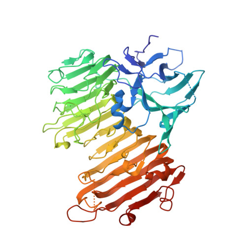
    N-glycosylation has been shown to be important for the stability of some glycoproteins. Isopullulanase (IPU), a polysaccharide-hydrolyzing enzyme, is a highly N-glycosylated protein, and IPU deglycosylation results in a decrease in thermostability. To investigate the function of N-glycan in IPU, we focused on an N-glycosylated residue located in the vicinity of the active site, Asn448. The thermostabilities of three IPU variants, Y440A, N448A and S450A, were 0.5-8.4°C lower than the wild-type enzyme. The crystal structure of endoglycosidase H (Endo H)-treated N448A variant was determined. There are four IPU molecules, Mol-A, B, C and D, in the asymmetric unit. The conformation of a loop composed of amino acid residues 435-455 in Mol-C was identical to wild-type IPU, whereas the conformations of this loop in Mol-A, Mol-B and Mol-D were different from each other. These results suggest that the Asn448 side chain is primarily important for the stability of IPU. Our results indicate that mutation of only N-glycosylated Asn residue may lead to incorrect conclusion for the evaluation of the function of N-glycan. Usually, the structures of N-glycosylation sites form an extended configuration in IPU; however, the Asn448 site had an atypical structure that lacked this configuration.


  • Organizational Affiliation
    • Department of Applied Biological Science, Tokyo University of Agriculture and Technology, 3-5-8 Saiwai-cho, Fuchu, Tokyo 183-8509, Japan.

Macromolecule Content 

  • Total Structure Weight: 247.75 kDa 
  • Atom Count: 17,816 
  • Modeled Residue Count: 2,159 
  • Deposited Residue Count: 2,196 
  • Unique protein chains: 1

Macromolecules

Find similar proteins by:|  3D Structure
Entity ID: 1
MoleculeChains  Sequence LengthOrganismDetailsImage
Isopullulanase
A, B, C, D
549Aspergillus nigerMutation(s): 1 
Gene Names: ipuA
EC: 3.2.1.57
UniProt
Find proteins for O00105 (Aspergillus niger)
Explore O00105 
Go to UniProtKB:  O00105
Entity Groups
Sequence Clusters30% Identity50% Identity70% Identity90% Identity95% Identity100% Identity
UniProt GroupO00105
Glycosylation
Glycosylation Sites: 8
Sequence Annotations
Expand
Reference Sequence

Oligosaccharides

Help  
Entity ID: 2
MoleculeChains Length2D Diagram GlycosylationD Interactions
alpha-D-glucopyranose-(1-6)-beta-D-glucopyranose
E
2N/A
Glycosylation Resources
GlyTouCan: G45624LJ
GlyCosmos: G45624LJ
Entity ID: 3
MoleculeChains Length2D Diagram GlycosylationD Interactions
alpha-D-glucopyranose-(1-4)-alpha-D-glucopyranose
F
2N/A
Glycosylation Resources
GlyTouCan: G07411ON
GlyCosmos: G07411ON

Small Molecules

Ligands 1 Unique
IDChains Name / Formula / InChI Key2D Diagram3D Interactions
NAG

Query on NAG


Download:Ideal Coordinates CCD File
AA [auth C]
BA [auth C]
CA [auth C]
DA [auth C]
EA [auth D]
AA [auth C],
BA [auth C],
CA [auth C],
DA [auth C],
EA [auth D],
FA [auth D],
G [auth A],
GA [auth D],
H [auth A],
HA [auth D],
I [auth A],
IA [auth D],
J [auth A],
JA [auth D],
K [auth A],
KA [auth D],
L [auth A],
LA [auth D],
M [auth A],
N [auth A],
O [auth B],
P [auth B],
Q [auth B],
R [auth B],
S [auth B],
T [auth B],
U [auth B],
V [auth C],
W [auth C],
X [auth C],
Y [auth C],
Z [auth C]
2-acetamido-2-deoxy-beta-D-glucopyranose
C8 H15 N O6
OVRNDRQMDRJTHS-FMDGEEDCSA-N

Biologically Interesting Molecules (External Reference) 

1 Unique

Experimental Data & Validation

Experimental Data

  • Method: X-RAY DIFFRACTION
  • Resolution: 2.20 Å
  • R-Value Free:  0.224 (Depositor), 0.226 (DCC) 
  • R-Value Work:  0.183 (Depositor), 0.191 (DCC) 
  • R-Value Observed: 0.185 (Depositor) 
Space Group: P 1 21 1
Unit Cell:
Length ( Å )Angle ( ˚ )
a = 96.4α = 90
b = 108.1β = 103.9
c = 116γ = 90
Software Package:
Software NamePurpose
ADSCdata collection
MOLREPphasing
REFMACrefinement
MOSFLMdata reduction
SCALAdata scaling

Structure Validation

View Full Validation Report


Ligand Structure Quality Assessment 


Entry History 

Deposition Data

Revision History  (Full details and data files)

  • Version 1.0: 2014-11-12
    Type: Initial release
  • Version 1.1: 2015-04-15
    Changes: Database references
  • Version 2.0: 2020-07-29
    Type: Remediation
    Reason: Carbohydrate remediation
    Changes: Atomic model, Data collection, Database references, Derived calculations, Non-polymer description, Structure summary
  • Version 2.1: 2023-11-08
    Changes: Data collection, Database references, Refinement description, Structure summary
  • Version 2.2: 2024-11-06
    Changes: Structure summary