3WSO | pdb_00003wso

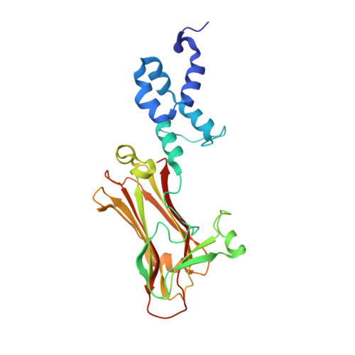
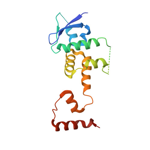
Crystal structure of the Skp1-FBG3 complex


Experimental Data Snapshot

  • Method: X-RAY DIFFRACTION
  • Resolution: 2.60 Å
  • R-Value Free: 
    0.266 (Depositor), 0.280 (DCC) 
  • R-Value Work: 
    0.205 (Depositor) 
  • R-Value Observed: 
    0.208 (Depositor) 

Starting Model: experimental
View more details

wwPDB Validation 3D Report Full Report

Validation slider image for 3WSO

This is version 1.3 of the entry. See complete history. 

Literature

The Structural Differences between a Glycoprotein Specific F-Box Protein Fbs1 and Its Homologous Protein FBG3

Kumanomidou, T., Nishio, K., Takagi, K., Nakagawa, T., Suzuki, A., Yamane, T., Tokunaga, F., Iwai, K., Murakami, A., Yoshida, Y., Tanaka, K., Mizushima, T.

(2015) PLoS One 10: e0140366-e0140366

  • DOI: https://doi.org/10.1371/journal.pone.0140366
  • Primary Citation Related Structures: 
    3WSO

  • PubMed Abstract: 

    The Skp1-Cul1-F-box protein (SCF) complex catalyzes protein ubiquitination in diverse cellular processes and is one of the best-characterized ubiquitin ligases. F-box proteins determine the substrate specificities of SCF ubiquitin ligases. Among these, Fbs1/FBG1/FBXO2, Fbs2/FBG2/FBXO6, and Fbs3/FBG5/FBXO27 recognize the N-glycans of glycoproteins, whereas FBG3/FBXO44 has no sugar-binding activity, despite the high sequence homology and conservation of the residues necessary for oligosaccharide binding between Fbs1-3 and FBG3. Here we determined the crystal structure of the Skp1-FBG3 complex at a resolution of 2.6 Å. The substrate-binding domain of FBG3 is composed of a 10-stranded antiparallel β-sandwich with three helices. Although the overall structure of FBG3 is similar to that of Fbs1, the residues that form the Fbs1 carbohydrate-binding pocket failed to be superposed with the corresponding residues of FBG3. Structure-based mutational analysis shows that distinct hydrogen bond networks of four FBG3 loops, i.e., β2-β3, β5-β6, β7-β8, and β9-β10, prevent the formation of the carbohydrate-binding pocket shown in Fbs1.


  • Organizational Affiliation: 
    • Department of Biotechnology, Graduate School of Engineering, Nagoya University, Chikusa-ku, Nagoya, Japan.

Macromolecule Content 

  • Total Structure Weight: 48.85 kDa 
  • Atom Count: 3,221 
  • Modeled Residue Count: 391 
  • Deposited Residue Count: 421 
  • Unique protein chains: 2

Macromolecules

Find similar proteins by:|  3D Structure
Entity ID: 1
MoleculeChains  Sequence LengthOrganismDetailsImage
F-box only protein 44255Homo sapiensMutation(s): 0 
Gene Names: FBXO44, FBG3, FBX30, FBX44, FBX6A, FBXO6A
UniProt & NIH Common Fund Data Resources
Find proteins for Q9H4M3 (Homo sapiens)
Explore Q9H4M3 
Go to UniProtKB:  Q9H4M3
PHAROS:  Q9H4M3
GTEx:  ENSG00000132879 
Entity Groups
Sequence Clusters30% Identity50% Identity70% Identity90% Identity95% Identity100% Identity
UniProt GroupQ9H4M3
Sequence Annotations
Expand
Reference Sequence
Find similar proteins by:|  3D Structure
Entity ID: 2
MoleculeChains  Sequence LengthOrganismDetailsImage
S-phase kinase-associated protein 1166Homo sapiensMutation(s): 0 
Gene Names: SKP1, EMC19, OCP2, SKP1A, TCEB1L
UniProt & NIH Common Fund Data Resources
Find proteins for P63208 (Homo sapiens)
Explore P63208 
Go to UniProtKB:  P63208
PHAROS:  P63208
GTEx:  ENSG00000113558 
Entity Groups
Sequence Clusters30% Identity50% Identity70% Identity90% Identity95% Identity100% Identity
UniProt GroupP63208
Sequence Annotations
Expand
Reference Sequence

Experimental Data & Validation

Experimental Data

  • Method: X-RAY DIFFRACTION
  • Resolution: 2.60 Å
  • R-Value Free:  0.266 (Depositor), 0.280 (DCC) 
  • R-Value Work:  0.205 (Depositor) 
  • R-Value Observed: 0.208 (Depositor) 
Space Group: P 21 21 21
Unit Cell:
Length ( Å )Angle ( ˚ )
a = 34.12α = 90
b = 76.594β = 90
c = 193.939γ = 90
Software Package:
Software NamePurpose
MOLREPphasing
REFMACrefinement
DENZOdata reduction
SCALEPACKdata scaling

Structure Validation

View Full Validation Report


Entry History 

Revision History  (Full details and data files)

  • Version 1.0: 2015-03-25
    Type: Initial release
  • Version 1.1: 2015-10-28
    Changes: Database references
  • Version 1.2: 2015-11-11
    Changes: Other
  • Version 1.3: 2023-11-08
    Changes: Data collection, Database references, Refinement description