3WA2 | pdb_00003wa2

High resolution crystal structure of copper amine oxidase from arthrobacter globiformis


Experimental Data Snapshot

  • Method: X-RAY DIFFRACTION
  • Resolution: 1.08 Å
  • R-Value Free: 
    0.150 (Depositor) 
  • R-Value Work: 
    0.130 (Depositor) 
  • R-Value Observed: 
    0.131 (Depositor) 

Starting Model: experimental
View more details

wwPDB Validation 3D Report Full Report

Validation slider image for 3WA2

This is version 1.3 of the entry. See complete history

Literature

High-resolution crystal structure of copper amine oxidase from Arthrobacter globiformis: assignment of bound diatomic molecules as O2

Murakawa, T.Hayashi, H.Sunami, T.Kurihara, K.Tamada, T.Kuroki, R.Suzuki, M.Tanizawa, K.Okajima, T.

(2013) Acta Crystallogr D Biol Crystallogr 69: 2483-2494

  • DOI: https://doi.org/10.1107/S0907444913023196
  • Primary Citation Related Structures: 
    3WA2, 3WA3

  • PubMed Abstract: 

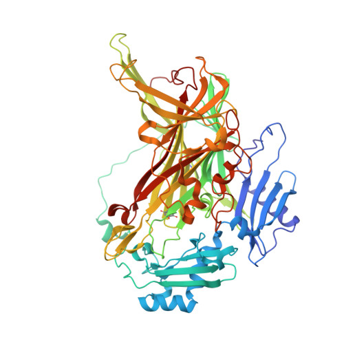
    The crystal structure of a copper amine oxidase from Arthrobacter globiformis was determined at 1.08 Å resolution with the use of low-molecular-weight polyethylene glycol (LMW PEG; average molecular weight ∼200) as a cryoprotectant. The final crystallographic R factor and Rfree were 13.0 and 15.0%, respectively. Several molecules of LMW PEG were found to occupy cavities in the protein interior, including the active site, which resulted in a marked reduction in the overall B factor and consequently led to a subatomic resolution structure for a relatively large protein with a monomer molecular weight of ∼70,000. About 40% of the presumed H atoms were observed as clear electron densities in the Fo - Fc difference map. Multiple minor conformers were also identified for many residues. Anisotropic displacement fluctuations were evaluated in the active site, which contains a post-translationally derived quinone cofactor and a Cu atom. Furthermore, diatomic molecules, most likely to be molecular oxygen, are bound to the protein, one of which is located in a region that had previously been proposed as an entry route for the dioxygen substrate from the central cavity of the dimer interface to the active site.


  • Organizational Affiliation
    • Department of Biochemistry, Osaka Medical College, 2-7 Daigakumachi, Takatsuki, Osaka 569-8686, Japan.

Macromolecule Content 

  • Total Structure Weight: 72.35 kDa 
  • Atom Count: 6,497 
  • Modeled Residue Count: 621 
  • Deposited Residue Count: 621 
  • Unique protein chains: 1

Macromolecules

Find similar proteins by:|  3D Structure
Entity ID: 1
MoleculeChains  Sequence LengthOrganismDetailsImage
Phenylethylamine oxidaseA [auth X]621Arthrobacter globiformisMutation(s): 0 
EC: 1.4.3.21
UniProt
Find proteins for P46881 (Arthrobacter globiformis)
Explore P46881 
Go to UniProtKB:  P46881
Entity Groups
Sequence Clusters30% Identity50% Identity70% Identity90% Identity95% Identity100% Identity
UniProt GroupP46881
Sequence Annotations
Expand
Reference Sequence

Small Molecules

Ligands 8 Unique
IDChains Name / Formula / InChI Key2D Diagram3D Interactions
1PE

Query on 1PE


Download:Ideal Coordinates CCD File
EA [auth X],
FA [auth X]
PENTAETHYLENE GLYCOL
C10 H22 O6
JLFNLZLINWHATN-UHFFFAOYSA-N
PG4

Query on PG4


Download:Ideal Coordinates CCD File
DA [auth X]TETRAETHYLENE GLYCOL
C8 H18 O5
UWHCKJMYHZGTIT-UHFFFAOYSA-N
PGE

Query on PGE


Download:Ideal Coordinates CCD File
AA [auth X],
BA [auth X],
CA [auth X],
Y [auth X],
Z [auth X]
TRIETHYLENE GLYCOL
C6 H14 O4
ZIBGPFATKBEMQZ-UHFFFAOYSA-N
PEG

Query on PEG


Download:Ideal Coordinates CCD File
P [auth X]
Q [auth X]
R [auth X]
S [auth X]
T [auth X]
P [auth X],
Q [auth X],
R [auth X],
S [auth X],
T [auth X],
U [auth X],
V [auth X],
W [auth X],
X
DI(HYDROXYETHYL)ETHER
C4 H10 O3
MTHSVFCYNBDYFN-UHFFFAOYSA-N
CU

Query on CU


Download:Ideal Coordinates CCD File
B [auth X]COPPER (II) ION
Cu
JPVYNHNXODAKFH-UHFFFAOYSA-N
EDO

Query on EDO


Download:Ideal Coordinates CCD File
D [auth X]
E [auth X]
F [auth X]
G [auth X]
H [auth X]
D [auth X],
E [auth X],
F [auth X],
G [auth X],
H [auth X],
I [auth X],
J [auth X],
K [auth X],
L [auth X],
M [auth X],
N [auth X],
O [auth X]
1,2-ETHANEDIOL
C2 H6 O2
LYCAIKOWRPUZTN-UHFFFAOYSA-N
OXY

Query on OXY


Download:Ideal Coordinates CCD File
GA [auth X],
HA [auth X],
IA [auth X],
JA [auth X]
OXYGEN MOLECULE
O2
MYMOFIZGZYHOMD-UHFFFAOYSA-N
NA

Query on NA


Download:Ideal Coordinates CCD File
C [auth X]SODIUM ION
Na
FKNQFGJONOIPTF-UHFFFAOYSA-N
Modified Residues  1 Unique
IDChains TypeFormula2D DiagramParent
TPQ
Query on TPQ
A [auth X]L-PEPTIDE LINKINGC9 H9 N O5TYR

Experimental Data & Validation

Experimental Data

  • Method: X-RAY DIFFRACTION
  • Resolution: 1.08 Å
  • R-Value Free:  0.150 (Depositor) 
  • R-Value Work:  0.130 (Depositor) 
  • R-Value Observed: 0.131 (Depositor) 
Space Group: C 1 2 1
Unit Cell:
Length ( Å )Angle ( ˚ )
a = 157.745α = 90
b = 62.379β = 112.1
c = 92.076γ = 90
Software Package:
Software NamePurpose
DENZOdata reduction
SCALEPACKdata scaling
PHENIXrefinement
PDB_EXTRACTdata extraction
HKL-2000data collection

Structure Validation

View Full Validation Report


Entry History 

Deposition Data

Revision History  (Full details and data files)

  • Version 1.0: 2013-09-11
    Type: Initial release
  • Version 1.1: 2014-01-01
    Changes: Database references
  • Version 1.2: 2017-11-22
    Changes: Refinement description
  • Version 1.3: 2023-11-08
    Changes: Data collection, Database references, Derived calculations, Refinement description