High-resolution crystal structure of copper amine oxidase from Arthrobacter globiformis: assignment of bound diatomic molecules as O2
Murakawa, T., Hayashi, H., Sunami, T., Kurihara, K., Tamada, T., Kuroki, R., Suzuki, M., Tanizawa, K., Okajima, T.(2013) Acta Crystallogr D Biol Crystallogr 69: 2483-2494
- PubMed: 24311589
- DOI: https://doi.org/10.1107/S0907444913023196
- Primary Citation of Related Structures:
3WA2, 3WA3 - PubMed Abstract:
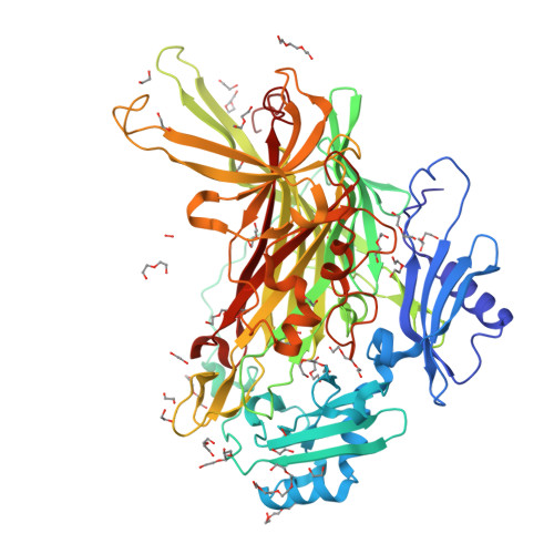
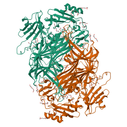
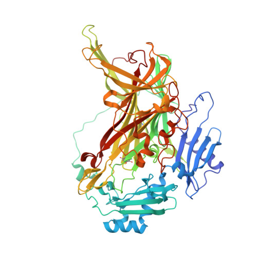
The crystal structure of a copper amine oxidase from Arthrobacter globiformis was determined at 1.08 Å resolution with the use of low-molecular-weight polyethylene glycol (LMW PEG; average molecular weight ∼200) as a cryoprotectant. The final crystallographic R factor and Rfree were 13.0 and 15.0%, respectively. Several molecules of LMW PEG were found to occupy cavities in the protein interior, including the active site, which resulted in a marked reduction in the overall B factor and consequently led to a subatomic resolution structure for a relatively large protein with a monomer molecular weight of ∼70,000. About 40% of the presumed H atoms were observed as clear electron densities in the Fo - Fc difference map. Multiple minor conformers were also identified for many residues. Anisotropic displacement fluctuations were evaluated in the active site, which contains a post-translationally derived quinone cofactor and a Cu atom. Furthermore, diatomic molecules, most likely to be molecular oxygen, are bound to the protein, one of which is located in a region that had previously been proposed as an entry route for the dioxygen substrate from the central cavity of the dimer interface to the active site.
Organizational Affiliation:
Department of Biochemistry, Osaka Medical College, 2-7 Daigakumachi, Takatsuki, Osaka 569-8686, Japan.


