3VPP | pdb_00003vpp

Crystal Structure of the Human CLEC9A C-type Lectin-Like Domain


Experimental Data Snapshot

  • Method: X-RAY DIFFRACTION
  • Resolution: 1.64 Å
  • R-Value Free: 
    0.227 (Depositor), 0.226 (DCC) 
  • R-Value Work: 
    0.189 (Depositor), 0.189 (DCC) 
  • R-Value Observed: 
    0.191 (Depositor) 

Starting Model: experimental
View more details

wwPDB Validation 3D Report Full Report

Validation slider image for 3VPP

This is version 1.5 of the entry. See complete history

Literature

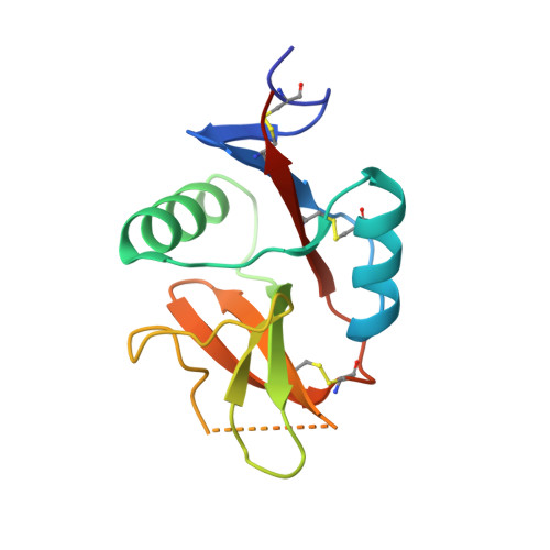
The dendritic cell receptor Clec9A binds damaged cells via exposed actin filaments.

Zhang, J.G.Czabotar, P.E.Policheni, A.N.Caminschi, I.Wan, S.S.Kitsoulis, S.Tullett, K.M.Robin, A.Y.Brammananth, R.van Delft, M.F.Lu, J.O'Reilly, L.A.Josefsson, E.C.Kile, B.T.Chin, W.J.Mintern, J.D.Olshina, M.A.Wong, W.Baum, J.Wright, M.D.Huang, D.C.Mohandas, N.Coppel, R.L.Colman, P.M.Nicola, N.A.Shortman, K.Lahoud, M.H.

(2012) Immunity 36: 646-657

  • DOI: https://doi.org/10.1016/j.immuni.2012.03.009
  • Primary Citation Related Structures: 
    3VPP

  • PubMed Abstract: 

    The immune system must distinguish viable cells from cells damaged by physical and infective processes. The damaged cell-recognition molecule Clec9A is expressed on the surface of the mouse and human dendritic cell subsets specialized for the uptake and processing of material from dead cells. Clec9A recognizes a conserved component within nucleated and nonnucleated cells, exposed when cell membranes are damaged. We have identified this Clec9A ligand as a filamentous form of actin in association with particular actin-binding domains of cytoskeletal proteins. We have determined the crystal structure of the human CLEC9A C-type lectin domain and propose a functional dimeric structure with conserved tryptophans in the ligand recognition site. Mutation of these residues ablated CLEC9A binding to damaged cells and to the isolated ligand complexes. We propose that Clec9A provides targeted recruitment of the adaptive immune system during infection and can also be utilized to enhance immune responses generated by vaccines.


  • Organizational Affiliation
    • The Walter and Eliza Hall Institute of Medical Research, Parkville, Victoria 3052, Australia.

Macromolecule Content 

  • Total Structure Weight: 30.2 kDa 
  • Atom Count: 2,292 
  • Modeled Residue Count: 243 
  • Deposited Residue Count: 264 
  • Unique protein chains: 1

Macromolecules

Find similar proteins by:|  3D Structure
Entity ID: 1
MoleculeChains  Sequence LengthOrganismDetailsImage
C-type lectin domain family 9 member A
A, B
132Homo sapiensMutation(s): 1 
Gene Names: CLEC9AUNQ9341/PRO34046
UniProt & NIH Common Fund Data Resources
Find proteins for Q6UXN8 (Homo sapiens)
Explore Q6UXN8 
Go to UniProtKB:  Q6UXN8
GTEx:  ENSG00000197992 
Entity Groups
Sequence Clusters30% Identity50% Identity70% Identity90% Identity95% Identity100% Identity
UniProt GroupQ6UXN8
Sequence Annotations
Expand
Reference Sequence

Experimental Data & Validation

Experimental Data

  • Method: X-RAY DIFFRACTION
  • Resolution: 1.64 Å
  • R-Value Free:  0.227 (Depositor), 0.226 (DCC) 
  • R-Value Work:  0.189 (Depositor), 0.189 (DCC) 
  • R-Value Observed: 0.191 (Depositor) 
Space Group: P 1 21 1
Unit Cell:
Length ( Å )Angle ( ˚ )
a = 42.17α = 90
b = 53.824β = 109.35
c = 57.446γ = 90
Software Package:
Software NamePurpose
Blu-Icedata collection
PHASERphasing
PHENIXrefinement
XDSdata reduction
XDSdata scaling

Structure Validation

View Full Validation Report


Entry History 

Deposition Data

Revision History  (Full details and data files)

  • Version 1.0: 2012-04-25
    Type: Initial release
  • Version 1.1: 2013-07-31
    Changes: Database references
  • Version 1.2: 2014-04-16
    Changes: Experimental preparation
  • Version 1.3: 2021-04-28
    Changes: Database references, Derived calculations, Source and taxonomy
  • Version 1.4: 2023-11-08
    Changes: Data collection, Database references, Refinement description
  • Version 1.5: 2024-10-30
    Changes: Structure summary