3VEP | pdb_00003vep

Crystal structure of SigD4 in complex with its negative regulator RsdA


Experimental Data Snapshot

  • Method: X-RAY DIFFRACTION
  • Resolution: 2.50 Å
  • R-Value Free: 
    0.287 (Depositor), 0.269 (DCC) 
  • R-Value Work: 
    0.243 (Depositor), 0.252 (DCC) 
  • R-Value Observed: 
    0.245 (Depositor) 

wwPDB Validation 3D Report Full Report

Validation slider image for 3VEP

This is version 1.2 of the entry. See complete history

Literature

Mycobacterium tuberculosis RsdA provides a conformational rationale for selective regulation of sigma-factor activity by proteolysis

Jaiswal, R.K.Prabha, T.S.Manjeera, G.Gopal, B.

(2013) Nucleic Acids Res 41: 3414-3423

  • DOI: https://doi.org/10.1093/nar/gks1468
  • Primary Citation Related Structures: 
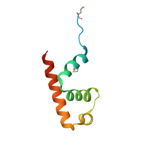
    3VEP, 3VFZ

  • PubMed Abstract: 

    The relative levels of different σ factors dictate the expression profile of a bacterium. Extracytoplasmic function σ factors synchronize the transcriptional profile with environmental conditions. The cellular concentration of free extracytoplasmic function σ factors is regulated by the localization of this protein in a σ/anti-σ complex. Anti-σ factors are multi-domain proteins with a receptor to sense environmental stimuli and a conserved anti-σ domain (ASD) that binds a σ factor. Here we describe the structure of Mycobacterium tuberculosis anti-σ(D) (RsdA) in complex with the -35 promoter binding domain of σ(D) (σ(D)4). We note distinct conformational features that enable the release of σ(D) by the selective proteolysis of the ASD in RsdA. The structural and biochemical features of the σ(D)/RsdA complex provide a basis to reconcile diverse regulatory mechanisms that govern σ/anti-σ interactions despite high overall structural similarity. Multiple regulatory mechanisms embedded in an ASD scaffold thus provide an elegant route to rapidly re-engineer the expression profile of a bacterium in response to an environmental stimulus.


  • Organizational Affiliation
    • Molecular Biophysics Unit, Indian Institute of Science, Bangalore 560 012, India.

Macromolecule Content 

  • Total Structure Weight: 74.82 kDa 
  • Atom Count: 3,680 
  • Modeled Residue Count: 466 
  • Deposited Residue Count: 664 
  • Unique protein chains: 2

Macromolecules

Find similar proteins by:|  3D Structure
Entity ID: 1
MoleculeChains  Sequence LengthOrganismDetailsImage
Uncharacterized protein Rv3413c/MT3522A [auth X],
C,
E [auth G],
G [auth J]
80Mycobacterium tuberculosisMutation(s): 0 
Gene Names: Rv3413c
UniProt
Find proteins for P9WJ71 (Mycobacterium tuberculosis (strain ATCC 25618 / H37Rv))
Explore P9WJ71 
Go to UniProtKB:  P9WJ71
Entity Groups
Sequence Clusters30% Identity50% Identity70% Identity90% Identity95% Identity100% Identity
UniProt GroupP9WJ71
Sequence Annotations
Expand
Reference Sequence
Find similar proteins by:|  3D Structure
Entity ID: 2
MoleculeChains  Sequence LengthOrganismDetailsImage
Probable RNA polymerase sigma-D factorB [auth D],
D [auth A],
F [auth E],
H
86Mycobacterium tuberculosisMutation(s): 0 
Gene Names: sigDRv3414c
UniProt
Find proteins for P9WGG9 (Mycobacterium tuberculosis (strain ATCC 25618 / H37Rv))
Explore P9WGG9 
Go to UniProtKB:  P9WGG9
Entity Groups
Sequence Clusters30% Identity50% Identity70% Identity90% Identity95% Identity100% Identity
UniProt GroupP9WGG9
Sequence Annotations
Expand
Reference Sequence

Small Molecules

Ligands 1 Unique
IDChains Name / Formula / InChI Key2D Diagram3D Interactions
SO4

Query on SO4


Download:Ideal Coordinates CCD File
I [auth X]
J [auth D]
K [auth D]
L [auth C]
M [auth A]
I [auth X],
J [auth D],
K [auth D],
L [auth C],
M [auth A],
N [auth A],
O [auth E],
P [auth E],
Q [auth E],
R [auth H],
S [auth H],
T [auth H],
U [auth H]
SULFATE ION
O4 S
QAOWNCQODCNURD-UHFFFAOYSA-L
Modified Residues  1 Unique
IDChains TypeFormula2D DiagramParent
MSE
Query on MSE
A [auth X],
C,
E [auth G],
G [auth J]
L-PEPTIDE LINKINGC5 H11 N O2 SeMET

Experimental Data & Validation

Experimental Data

  • Method: X-RAY DIFFRACTION
  • Resolution: 2.50 Å
  • R-Value Free:  0.287 (Depositor), 0.269 (DCC) 
  • R-Value Work:  0.243 (Depositor), 0.252 (DCC) 
  • R-Value Observed: 0.245 (Depositor) 
Space Group: C 1 2 1
Unit Cell:
Length ( Å )Angle ( ˚ )
a = 99.74α = 90
b = 110.72β = 133
c = 73.13γ = 90
Software Package:
Software NamePurpose
HKL-2000data collection
SOLVEphasing
PHENIXrefinement
MOSFLMdata reduction
SCALAdata scaling

Structure Validation

View Full Validation Report


Entry History 

Deposition Data

Revision History  (Full details and data files)

  • Version 1.0: 2013-02-13
    Type: Initial release
  • Version 1.1: 2013-10-09
    Changes: Database references
  • Version 1.2: 2024-10-09
    Changes: Data collection, Database references, Derived calculations, Structure summary