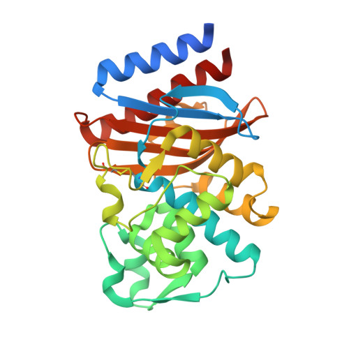
3TSG

Crystal structure of GES-14


Experimental Data Snapshot

  • Method: X-RAY DIFFRACTION
  • Resolution: 1.90 Å
  • R-Value Free: 0.205 
  • R-Value Work: 0.163 
  • R-Value Observed: 0.165 

Starting Model: experimental
View more details

wwPDB Validation   3D Report Full Report


This is version 1.3 of the entry. See complete history


Literature

Kinetic and crystallographic studies of extended-spectrum GES-11, GES-12, and GES-14 beta-lactamases.

Delbruck, H.Bogaerts, P.Kupper, M.B.Rezende de Castro, R.Bennink, S.Glupczynski, Y.Galleni, M.Hoffmann, K.M.Bebrone, C.

(2012) Antimicrob Agents Chemother 56: 5618-5625

  • DOI: https://doi.org/10.1128/AAC.01272-12
  • Primary Citation of Related Structures:  
    3TSG, 3V3R

  • PubMed Abstract: 

    GES-1 is a class A extended-spectrum β-lactamase conferring resistance to penicillins, narrow- and expanded-spectrum cephalosporins, and ceftazidime. However, GES-1 poorly hydrolyzes aztreonam and cephamycins and exhibits very low k(cat) values for carbapenems. Twenty-two GES variants have been discovered thus far, differing from each other by 1 to 3 amino acid substitutions that affect substrate specificity. GES-11 possesses a Gly243Ala substitution which seems to confer to this variant an increased activity against aztreonam and ceftazidime. GES-12 differs from GES-11 by a single Thr237Ala substitution, while GES-14 differs from GES-11 by the Gly170Ser mutation, which is known to confer increased carbapenemase activity. GES-11 and GES-12 were kinetically characterized and compared to GES-1 and GES-14. Purified GES-11 and GES-12 showed strong activities against most tested β-lactams, with the exception of temocillin, cefoxitin, and carbapenems. Both variants showed a significantly increased rate of hydrolysis of cefotaxime, ceftazidime, and aztreonam. On the other hand, GES-11 and GES-12 (and GES-14) variants all containing Ala243 exhibited increased susceptibility to classical inhibitors. The crystallographic structures of the GES-11 and GES-14 β-lactamases were solved. The overall structures of GES-11 and GES-14 are similar to that of GES-1. The Gly243Ala substitution caused only subtle local rearrangements, notably in the typical carbapenemase disulfide bond. The active sites of GES-14 and GES-11 are very similar, with the Gly170Ser substitution leading only to the formation of additional hydrogen bonds of the Ser residue with hydrolytic water and the Glu166 residue.


  • Organizational Affiliation

    Institute of Molecular Biotechnology, RWTH-Aachen University, Aachen, Germany.


Macromolecules
Find similar proteins by:  (by identity cutoff)  |  3D Structure
Entity ID: 1
MoleculeChains Sequence LengthOrganismDetailsImage
Extended-spectrum beta-lactamase GES-14
A, B
287Acinetobacter baumanniiMutation(s): 0 
Gene Names: blaGES-14
EC: 3.5.2.6
UniProt
Find proteins for D3X610 (Acinetobacter baumannii)
Explore D3X610 
Go to UniProtKB:  D3X610
Entity Groups  
Sequence Clusters30% Identity50% Identity70% Identity90% Identity95% Identity100% Identity
UniProt GroupD3X610
Sequence Annotations
Expand
  • Reference Sequence
Small Molecules
Ligands 2 Unique
IDChains Name / Formula / InChI Key2D Diagram3D Interactions
IOD
Query on IOD

Download Ideal Coordinates CCD File 
AA [auth A]
AB [auth B]
BA [auth A]
BB [auth B]
C [auth A]
AA [auth A],
AB [auth B],
BA [auth A],
BB [auth B],
C [auth A],
CA [auth A],
CB [auth B],
D [auth A],
DA [auth A],
DB [auth B],
E [auth A],
EA [auth A],
EB [auth B],
F [auth A],
FA [auth A],
FB [auth B],
G [auth A],
GB [auth B],
H [auth A],
HB [auth B],
I [auth A],
IB [auth B],
J [auth A],
K [auth A],
KA [auth B],
L [auth A],
LA [auth B],
M [auth A],
MA [auth B],
N [auth A],
NA [auth B],
O [auth A],
OA [auth B],
P [auth A],
PA [auth B],
Q [auth A],
QA [auth B],
R [auth A],
RA [auth B],
S [auth A],
SA [auth B],
T [auth A],
TA [auth B],
U [auth A],
UA [auth B],
V [auth A],
VA [auth B],
W [auth A],
WA [auth B],
X [auth A],
XA [auth B],
Y [auth A],
YA [auth B],
Z [auth A],
ZA [auth B]
IODIDE ION
I
XMBWDFGMSWQBCA-UHFFFAOYSA-M
GOL
Query on GOL

Download Ideal Coordinates CCD File 
GA [auth A]
HA [auth A]
IA [auth A]
JA [auth A]
JB [auth B]
GA [auth A],
HA [auth A],
IA [auth A],
JA [auth A],
JB [auth B],
KB [auth B],
LB [auth B],
MB [auth B],
NB [auth B],
OB [auth B],
PB [auth B]
GLYCEROL
C3 H8 O3
PEDCQBHIVMGVHV-UHFFFAOYSA-N
Experimental Data & Validation

Experimental Data

  • Method: X-RAY DIFFRACTION
  • Resolution: 1.90 Å
  • R-Value Free: 0.205 
  • R-Value Work: 0.163 
  • R-Value Observed: 0.165 
  • Space Group: P 2 21 21
Unit Cell:
Length ( Å )Angle ( ˚ )
a = 70.745α = 90
b = 84.098β = 90
c = 85.026γ = 90
Software Package:
Software NamePurpose
MAR345dtbdata collection
PHASERphasing
REFMACrefinement
XDSdata reduction
SCALAdata scaling

Structure Validation

View Full Validation Report


Entry History 

Deposition Data

Revision History  (Full details and data files)

  • Version 1.0: 2012-09-26
    Type: Initial release
  • Version 1.1: 2013-06-19
    Changes: Database references
  • Version 1.2: 2023-09-13
    Changes: Data collection, Database references, Derived calculations, Refinement description
  • Version 1.3: 2024-10-09
    Changes: Structure summary