3V3R | pdb_00003v3r

Crystal Structure of GES-11


Experimental Data Snapshot

  • Method: X-RAY DIFFRACTION
  • Resolution: 1.90 Å
  • R-Value Free: 
    0.232 (Depositor), 0.233 (DCC) 
  • R-Value Work: 
    0.192 (Depositor), 0.197 (DCC) 
  • R-Value Observed: 
    0.194 (Depositor) 

Starting Model: experimental
View more details

wwPDB Validation 3D Report Full Report

Validation slider image for 3V3R

This is version 1.3 of the entry. See complete history

Literature

Kinetic and crystallographic studies of extended-spectrum GES-11, GES-12, and GES-14 beta-lactamases.

Delbruck, H.Bogaerts, P.Kupper, M.B.Rezende de Castro, R.Bennink, S.Glupczynski, Y.Galleni, M.Hoffmann, K.M.Bebrone, C.

(2012) Antimicrob Agents Chemother 56: 5618-5625

  • DOI: https://doi.org/10.1128/AAC.01272-12
  • Primary Citation Related Structures: 
    3TSG, 3V3R

  • PubMed Abstract: 

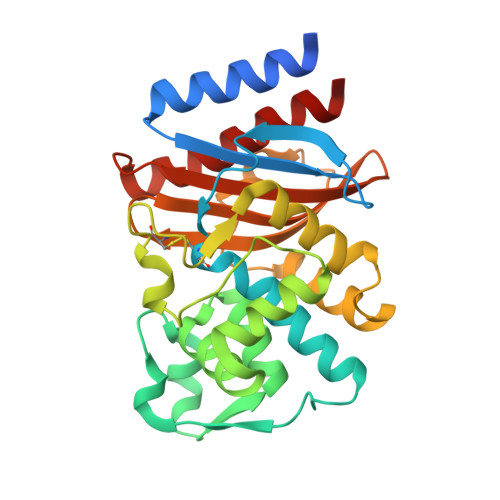
    GES-1 is a class A extended-spectrum β-lactamase conferring resistance to penicillins, narrow- and expanded-spectrum cephalosporins, and ceftazidime. However, GES-1 poorly hydrolyzes aztreonam and cephamycins and exhibits very low k(cat) values for carbapenems. Twenty-two GES variants have been discovered thus far, differing from each other by 1 to 3 amino acid substitutions that affect substrate specificity. GES-11 possesses a Gly243Ala substitution which seems to confer to this variant an increased activity against aztreonam and ceftazidime. GES-12 differs from GES-11 by a single Thr237Ala substitution, while GES-14 differs from GES-11 by the Gly170Ser mutation, which is known to confer increased carbapenemase activity. GES-11 and GES-12 were kinetically characterized and compared to GES-1 and GES-14. Purified GES-11 and GES-12 showed strong activities against most tested β-lactams, with the exception of temocillin, cefoxitin, and carbapenems. Both variants showed a significantly increased rate of hydrolysis of cefotaxime, ceftazidime, and aztreonam. On the other hand, GES-11 and GES-12 (and GES-14) variants all containing Ala243 exhibited increased susceptibility to classical inhibitors. The crystallographic structures of the GES-11 and GES-14 β-lactamases were solved. The overall structures of GES-11 and GES-14 are similar to that of GES-1. The Gly243Ala substitution caused only subtle local rearrangements, notably in the typical carbapenemase disulfide bond. The active sites of GES-14 and GES-11 are very similar, with the Gly170Ser substitution leading only to the formation of additional hydrogen bonds of the Ser residue with hydrolytic water and the Glu166 residue.


  • Organizational Affiliation
    • Institute of Molecular Biotechnology, RWTH-Aachen University, Aachen, Germany.

Macromolecule Content 

  • Total Structure Weight: 65.35 kDa 
  • Atom Count: 4,453 
  • Modeled Residue Count: 531 
  • Deposited Residue Count: 574 
  • Unique protein chains: 1

Macromolecules

Find similar proteins by:|  3D Structure
Entity ID: 1
MoleculeChains  Sequence LengthOrganismDetailsImage
Extended spectrum class A beta-lactamase GES-11
A, B
287Acinetobacter baumanniiMutation(s): 0 
Gene Names: blaGES-11GES11
EC: 3.5.2.6
UniProt
Find proteins for C5HUY1 (Acinetobacter baumannii)
Explore C5HUY1 
Go to UniProtKB:  C5HUY1
Entity Groups
Sequence Clusters30% Identity50% Identity70% Identity90% Identity95% Identity100% Identity
UniProt GroupC5HUY1
Sequence Annotations
Expand
Reference Sequence

Small Molecules

Ligands 2 Unique
IDChains Name / Formula / InChI Key2D Diagram3D Interactions
IOD

Query on IOD


Download:Ideal Coordinates CCD File
C [auth A]
D [auth A]
E [auth A]
F [auth A]
G [auth A]
C [auth A],
D [auth A],
E [auth A],
F [auth A],
G [auth A],
H [auth A],
I [auth A],
J [auth A],
K [auth A],
L [auth A],
M [auth A],
N [auth B],
O [auth B],
P [auth B],
Q [auth B],
R [auth B],
S [auth B],
T [auth B],
U [auth B],
V [auth B],
W [auth B],
X [auth B],
Y [auth B]
IODIDE ION
I
XMBWDFGMSWQBCA-UHFFFAOYSA-M
NA

Query on NA


Download:Ideal Coordinates CCD File
Z [auth B]SODIUM ION
Na
FKNQFGJONOIPTF-UHFFFAOYSA-N

Experimental Data & Validation

Experimental Data

  • Method: X-RAY DIFFRACTION
  • Resolution: 1.90 Å
  • R-Value Free:  0.232 (Depositor), 0.233 (DCC) 
  • R-Value Work:  0.192 (Depositor), 0.197 (DCC) 
  • R-Value Observed: 0.194 (Depositor) 
Space Group: P 2 21 21
Unit Cell:
Length ( Å )Angle ( ˚ )
a = 70.68α = 90
b = 85.24β = 90
c = 85.12γ = 90
Software Package:
Software NamePurpose
MAR345dtbdata collection
PHASERphasing
REFMACrefinement
XDSdata reduction
XDSdata scaling

Structure Validation

View Full Validation Report


Entry History 

Deposition Data

Revision History  (Full details and data files)

  • Version 1.0: 2012-09-26
    Type: Initial release
  • Version 1.1: 2013-06-19
    Changes: Database references
  • Version 1.2: 2023-09-13
    Changes: Data collection, Database references, Derived calculations, Refinement description
  • Version 1.3: 2024-10-09
    Changes: Structure summary