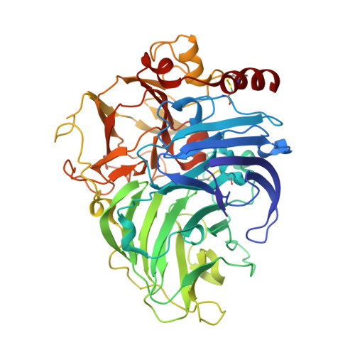
Reaction intermediates and redox state changes in a blue laccase from Steccherinum ochraceum observed by crystallographic high/low X-ray dose experiments.
Ferraroni, M., Matera, I., Chernykh, A., Kolomytseva, M., Golovleva, L.A., Scozzafava, A., Briganti, F.(2012) J Inorg Biochem 111: 203-209
- PubMed: 22341982
- DOI: https://doi.org/10.1016/j.jinorgbio.2012.01.011
- Primary Citation of Related Structures:
3T6V, 3T6W, 3T6X, 3T6Z, 3T71 - PubMed Abstract:
The crystal structure of a blue laccase from Steccherinum ochraceum has been solved at 2.0Å of resolution using a classic data acquisition from a single crystal. The overall structural features are typical of this class of enzymes, however, distances inside the trinuclear copper cluster are indicative of a reduction of the metal centers induced by free electrons produced during the X-ray data collection. UV-visible spectra collected during the X-ray exposure support the progressive reduction of the metal centers. In order to better detect the reduction progression steps in the trinuclear copper site, a multicrystal data collection strategy based on a systematic spread of the X-ray dose over many crystals has been employed. This approach is based on collecting multicrystal data sets, then combining the slices of the individual data sets experiencing the same radiation dose to obtain composite complete data sets at progressively higher doses. Applying this technique, we have been able to capture sequential frames of the enzyme during the metal centers and molecular oxygen reduction mechanism obtaining a three-dimensional movie of the X-ray-driven catalytic conversion of the molecular oxygen in the active site of laccase: first, the copper ions reduction, then the molecular oxygen binding and its reductive splitting, thus allowing to reconstruct the entire catalytic cycle for multicopper oxidases.
- Dipartimento di Chimica, Università di Firenze, Via della Lastruccia 3, Sesto Fiorentino, Florence, Italy.
Organizational Affiliation:


