3T6X | pdb_00003t6x

Crystal Structure of Steccherinum ochraceum Laccase obtained by multi-crystals composite data collection technique (20% dose)


Experimental Data Snapshot

  • Method: X-RAY DIFFRACTION
  • Resolution: 2.15 Å
  • R-Value Free: 
    0.236 (Depositor), 0.237 (DCC) 
  • R-Value Work: 
    0.176 (Depositor), 0.178 (DCC) 
  • R-Value Observed: 
    0.179 (Depositor) 

Starting Model: experimental
View more details

wwPDB Validation 3D Report Full Report

Validation slider image for 3T6X

Ligand Structure Quality Assessment 


This is version 2.2 of the entry. See complete history

Literature

Reaction intermediates and redox state changes in a blue laccase from Steccherinum ochraceum observed by crystallographic high/low X-ray dose experiments.

Ferraroni, M.Matera, I.Chernykh, A.Kolomytseva, M.Golovleva, L.A.Scozzafava, A.Briganti, F.

(2012) J Inorg Biochem 111: 203-209

  • DOI: https://doi.org/10.1016/j.jinorgbio.2012.01.011
  • Primary Citation Related Structures: 
    3T6V, 3T6W, 3T6X, 3T6Z, 3T71

  • PubMed Abstract: 

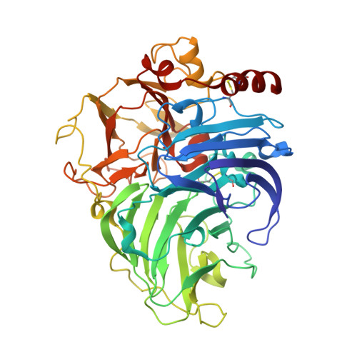
    The crystal structure of a blue laccase from Steccherinum ochraceum has been solved at 2.0Å of resolution using a classic data acquisition from a single crystal. The overall structural features are typical of this class of enzymes, however, distances inside the trinuclear copper cluster are indicative of a reduction of the metal centers induced by free electrons produced during the X-ray data collection. UV-visible spectra collected during the X-ray exposure support the progressive reduction of the metal centers. In order to better detect the reduction progression steps in the trinuclear copper site, a multicrystal data collection strategy based on a systematic spread of the X-ray dose over many crystals has been employed. This approach is based on collecting multicrystal data sets, then combining the slices of the individual data sets experiencing the same radiation dose to obtain composite complete data sets at progressively higher doses. Applying this technique, we have been able to capture sequential frames of the enzyme during the metal centers and molecular oxygen reduction mechanism obtaining a three-dimensional movie of the X-ray-driven catalytic conversion of the molecular oxygen in the active site of laccase: first, the copper ions reduction, then the molecular oxygen binding and its reductive splitting, thus allowing to reconstruct the entire catalytic cycle for multicopper oxidases.


  • Organizational Affiliation
    • Dipartimento di Chimica, Università di Firenze, Via della Lastruccia 3, Sesto Fiorentino, Florence, Italy.

Macromolecule Content 

  • Total Structure Weight: 163.1 kDa 
  • Atom Count: 12,916 
  • Modeled Residue Count: 1,485 
  • Deposited Residue Count: 1,485 
  • Unique protein chains: 1

Macromolecules

Find similar proteins by:|  3D Structure
Entity ID: 1
MoleculeChains  Sequence LengthOrganismDetailsImage
Laccase
A, B, C
495Steccherinum ochraceumMutation(s): 0 
EC: 1.10.3.2
UniProt
Find proteins for I1SB14 (Steccherinum ochraceum)
Explore I1SB14 
Go to UniProtKB:  I1SB14
Entity Groups
Sequence Clusters30% Identity50% Identity70% Identity90% Identity95% Identity100% Identity
UniProt GroupI1SB14
Glycosylation
Glycosylation Sites: 2
Sequence Annotations
Expand
Reference Sequence

Oligosaccharides

Help  
Entity ID: 2
MoleculeChains Length2D Diagram GlycosylationD Interactions
2-acetamido-2-deoxy-beta-D-glucopyranose-(1-4)-2-acetamido-2-deoxy-beta-D-glucopyranose
D, E, F, G, H
2N-Glycosylation
Glycosylation Resources
GlyTouCan: G42666HT
GlyCosmos: G42666HT
GlyGen: G42666HT

Small Molecules

Ligands 5 Unique
IDChains Name / Formula / InChI Key2D Diagram3D Interactions
NAG

Query on NAG


Download:Ideal Coordinates CCD File
LA [auth C]2-acetamido-2-deoxy-beta-D-glucopyranose
C8 H15 N O6
OVRNDRQMDRJTHS-FMDGEEDCSA-N
SO4

Query on SO4


Download:Ideal Coordinates CCD File
IA [auth C]
M [auth A]
N [auth A]
O [auth A]
W [auth B]
IA [auth C],
M [auth A],
N [auth A],
O [auth A],
W [auth B],
X [auth B],
Y [auth B]
SULFATE ION
O4 S
QAOWNCQODCNURD-UHFFFAOYSA-L
GOL

Query on GOL


Download:Ideal Coordinates CCD File
AA [auth B]
BA [auth B]
CA [auth B]
JA [auth C]
KA [auth C]
AA [auth B],
BA [auth B],
CA [auth B],
JA [auth C],
KA [auth C],
P [auth A],
Q [auth A],
Z [auth B]
GLYCEROL
C3 H8 O3
PEDCQBHIVMGVHV-UHFFFAOYSA-N
CU

Query on CU


Download:Ideal Coordinates CCD File
EA [auth C]
FA [auth C]
GA [auth C]
HA [auth C]
I [auth A]
EA [auth C],
FA [auth C],
GA [auth C],
HA [auth C],
I [auth A],
J [auth A],
K [auth A],
L [auth A],
S [auth B],
T [auth B],
U [auth B],
V [auth B]
COPPER (II) ION
Cu
JPVYNHNXODAKFH-UHFFFAOYSA-N
PER

Query on PER


Download:Ideal Coordinates CCD File
DA [auth B],
MA [auth C],
R [auth A]
PEROXIDE ION
O2
ANAIPYUSIMHBEL-UHFFFAOYSA-N

Experimental Data & Validation

Experimental Data

  • Method: X-RAY DIFFRACTION
  • Resolution: 2.15 Å
  • R-Value Free:  0.236 (Depositor), 0.237 (DCC) 
  • R-Value Work:  0.176 (Depositor), 0.178 (DCC) 
  • R-Value Observed: 0.179 (Depositor) 
Space Group: P 21 21 21
Unit Cell:
Length ( Å )Angle ( ˚ )
a = 73.51α = 90
b = 140.04β = 90
c = 172.98γ = 90
Software Package:
Software NamePurpose
XSCALEdata scaling
REFMACrefinement
PDB_EXTRACTdata extraction
MAR345dtbdata collection
XDSdata reduction

Structure Validation

View Full Validation Report


Ligand Structure Quality Assessment 


Entry History 

Deposition Data

Revision History  (Full details and data files)

  • Version 1.0: 2012-04-18
    Type: Initial release
  • Version 1.1: 2012-10-03
    Changes: Database references
  • Version 2.0: 2020-07-29
    Type: Remediation
    Reason: Carbohydrate remediation
    Changes: Advisory, Atomic model, Data collection, Derived calculations, Non-polymer description, Structure summary
  • Version 2.1: 2023-09-13
    Changes: Data collection, Database references, Derived calculations, Refinement description, Structure summary
  • Version 2.2: 2024-10-16
    Changes: Structure summary