3T5C | pdb_00003t5c

Crystal structure of N-terminal domain of FACL13 from Mycobacterium tuberculosis in different space group C2


Experimental Data Snapshot

  • Method: X-RAY DIFFRACTION
  • Resolution: 2.09 Å
  • R-Value Free: 
    0.263 (Depositor), 0.207 (DCC) 
  • R-Value Work: 
    0.208 (Depositor), 0.205 (DCC) 
  • R-Value Observed: 
    0.208 (Depositor) 

Starting Model: experimental
View more details

wwPDB Validation 3D Report Full Report

Validation slider image for 3T5C

This is version 1.2 of the entry. See complete history

Literature

Molecular basis of the functional divergence of fatty acyl-AMP ligase biosynthetic enzymes of Mycobacterium tuberculosis.

Goyal, A.Verma, P.Anandhakrishnan, M.Gokhale, R.S.Sankaranarayanan, R.

(2012) J Mol Biology 416: 221-238

  • DOI: https://doi.org/10.1016/j.jmb.2011.12.031
  • Primary Citation Related Structures: 
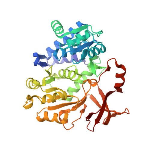
    3T5A, 3T5B, 3T5C

  • PubMed Abstract: 

    Activation of fatty acids as acyl-adenylates by fatty acyl-AMP ligases (FAALs) in Mycobacterium tuberculosis is a variant of a classical theme that involves formation of acyl-CoA (coenzyme A) by fatty acyl-CoA ligases (FACLs). Here, we show that FAALs and FACLs possess similar structural fold and substrate specificity determinants, and the key difference is the absence of a unique insertion sequence in FACL13 structure. A systematic analysis shows a conserved hydrophobic anchorage of the insertion motif across several FAALs. Strikingly, mutagenesis of two phenylalanine residues, which are part of the anchorage, to alanine converts FAAL32 to FACL32. This insertion-based in silico analysis suggests the presence of FAAL homologues in several other non-mycobacterial genomes including eukaryotes. The work presented here establishes an elegant mechanism wherein an insertion sequence drives the functional divergence of FAALs from canonical FACLs.


  • Organizational Affiliation
    • Centre for Cellular and Molecular Biology, Council of Scientific and Industrial Research, Hyderabad 500 007, India.

Macromolecule Content 

  • Total Structure Weight: 85.68 kDa 
  • Atom Count: 6,461 
  • Modeled Residue Count: 784 
  • Deposited Residue Count: 792 
  • Unique protein chains: 1

Macromolecules

Find similar proteins by:|  3D Structure
Entity ID: 1
MoleculeChains  Sequence LengthOrganismDetailsImage
PROBABLE CHAIN-FATTY-ACID-CoA LIGASE FADD13
A, B
396Mycobacterium tuberculosis H37RvMutation(s): 0 
Gene Names: fadD13MT3174Rv3089
EC: 6.2.1 (PDB Primary Data), 6.2.1.3 (UniProt)
UniProt
Find proteins for P9WQ37 (Mycobacterium tuberculosis (strain ATCC 25618 / H37Rv))
Explore P9WQ37 
Go to UniProtKB:  P9WQ37
Entity Groups
Sequence Clusters30% Identity50% Identity70% Identity90% Identity95% Identity100% Identity
UniProt GroupP9WQ37
Sequence Annotations
Expand
Reference Sequence

Experimental Data & Validation

Experimental Data

  • Method: X-RAY DIFFRACTION
  • Resolution: 2.09 Å
  • R-Value Free:  0.263 (Depositor), 0.207 (DCC) 
  • R-Value Work:  0.208 (Depositor), 0.205 (DCC) 
  • R-Value Observed: 0.208 (Depositor) 
Space Group: C 1 2 1
Unit Cell:
Length ( Å )Angle ( ˚ )
a = 129.228α = 90
b = 91.983β = 130.79
c = 84.412γ = 90
Software Package:
Software NamePurpose
MAR345dtbdata collection
MOLREPphasing
CNSrefinement
DENZOdata reduction
SCALEPACKdata scaling

Structure Validation

View Full Validation Report


Entry History 

Deposition Data

Revision History  (Full details and data files)

  • Version 1.0: 2012-01-25
    Type: Initial release
  • Version 1.1: 2013-07-10
    Changes: Database references
  • Version 1.2: 2023-11-01
    Changes: Data collection, Database references, Refinement description