3SO3 | pdb_00003so3

Structures of Fab-Protease Complexes Reveal a Highly Specific Non-Canonical Mechanism of Inhibition.


Experimental Data Snapshot

  • Method: X-RAY DIFFRACTION
  • Resolution: 2.10 Å
  • R-Value Free: 
    0.194 (Depositor), 0.203 (DCC) 
  • R-Value Work: 
    0.161 (Depositor), 0.172 (DCC) 
  • R-Value Observed: 
    0.162 (Depositor) 

Starting Model: experimental
View more details

wwPDB Validation 3D Report Full Report

Validation slider image for 3SO3

This is version 2.2 of the entry. See complete history

Literature

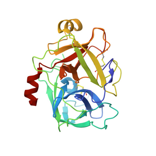
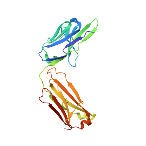
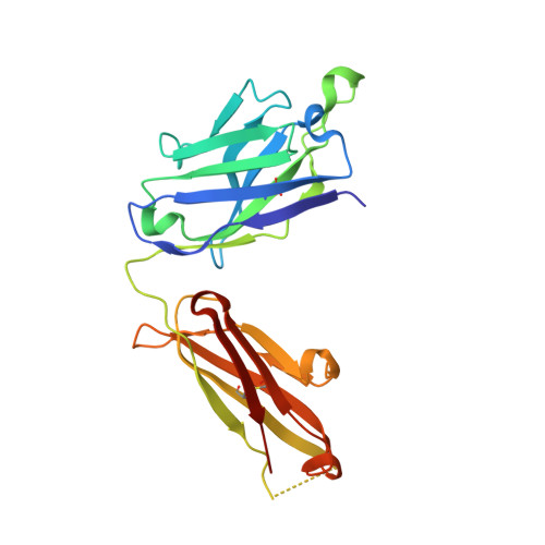
A reverse binding motif that contributes to specific protease inhibition by antibodies.

Schneider, E.L.Lee, M.S.Baharuddin, A.Goetz, D.H.Farady, C.J.Ward, M.Wang, C.I.Craik, C.S.

(2012) J Mol Biology 415: 699-715

  • DOI: https://doi.org/10.1016/j.jmb.2011.11.036
  • Primary Citation Related Structures: 
    3NPS, 3SO3

  • PubMed Abstract: 

    The type II transmembrane serine protease family consists of 18 closely related serine proteases that are implicated in multiple functions. To identify selective, inhibitory antibodies against one particular type II transmembrane serine protease, matriptase [MT-SP1 (membrane-type serine protease 1)], a phage display library was created with a natural repertoire of Fabs [fragment antigen binding (Fab)] from human naïve B cells. Fab A11 was identified with a 720 pM inhibition constant and high specificity for matriptase over other trypsin-fold serine proteases. A Trichoderma reesei system expressed A11 with a yield of ∼200 mg/L. The crystal structure of A11 in complex with matriptase has been determined and compared to the crystal structure of another antibody inhibitor (S4) in complex with matriptase. Previously discovered from a synthetic single-chain variable fragment library, S4 is also a highly selective and potent matriptase inhibitor. The crystal structures of the A11/matriptase and S4/matriptase complexes were solved to 2.1 Å and 1.5 Å, respectively. Although these antibodies, discovered from separate libraries, interact differently with the protease surface loops for their specificity, the structures reveal a similar novel mechanism of protease inhibition. Through the insertion of the H3 variable loop in a reverse orientation at the substrate-binding pocket, these antibodies bury a large surface area for potent inhibition and avoid proteolytic inactivation. This discovery highlights the critical role that the antibody scaffold plays in positioning loops to bind and inhibit protease function in a highly selective manner. Additionally, Fab A11 is a fully human antibody that specifically inhibits matriptase over other closely related proteases, suggesting that this approach could be useful for clinical applications.


  • Organizational Affiliation
    • Department of Pharmaceutical Chemistry, University of California, San Francisco, Genentech Hall, San Francisco, CA 94143-2280, USA.

Macromolecule Content 

  • Total Structure Weight: 74.33 kDa 
  • Atom Count: 5,605 
  • Modeled Residue Count: 676 
  • Deposited Residue Count: 686 
  • Unique protein chains: 3

Macromolecules

Find similar proteins by:|  3D Structure
Entity ID: 1
MoleculeChains  Sequence LengthOrganismDetailsImage
Suppressor of tumorigenicity 14 protein241Homo sapiensMutation(s): 1 
Gene Names: PRSS14SNC19ST14TADG15
EC: 3.4.21.109
UniProt & NIH Common Fund Data Resources
Find proteins for Q9Y5Y6 (Homo sapiens)
Explore Q9Y5Y6 
Go to UniProtKB:  Q9Y5Y6
PHAROS:  Q9Y5Y6
GTEx:  ENSG00000149418 
Entity Groups
Sequence Clusters30% Identity50% Identity70% Identity90% Identity95% Identity100% Identity
UniProt GroupQ9Y5Y6
Sequence Annotations
Expand
Reference Sequence
Find similar proteins by:|  3D Structure
Entity ID: 2
MoleculeChains  Sequence LengthOrganismDetailsImage
A11 FAB light chain217Homo sapiensMutation(s): 0 
Find similar proteins by:|  3D Structure
Entity ID: 3
MoleculeChains  Sequence LengthOrganismDetailsImage
A11 FAB heavy chain228Homo sapiensMutation(s): 0 

Oligosaccharides

Help  
Entity ID: 4
MoleculeChains Length2D Diagram GlycosylationD Interactions
beta-D-fructofuranose-(2-1)-alpha-D-glucopyranose
D
2N/A
Glycosylation Resources
GlyTouCan: G05551OP
GlyCosmos: G05551OP

Small Molecules

Ligands 1 Unique
IDChains Name / Formula / InChI Key2D Diagram3D Interactions
GOL

Query on GOL


Download:Ideal Coordinates CCD File
E [auth A]GLYCEROL
C3 H8 O3
PEDCQBHIVMGVHV-UHFFFAOYSA-N

Biologically Interesting Molecules (External Reference) 

1 Unique

Experimental Data & Validation

Experimental Data

  • Method: X-RAY DIFFRACTION
  • Resolution: 2.10 Å
  • R-Value Free:  0.194 (Depositor), 0.203 (DCC) 
  • R-Value Work:  0.161 (Depositor), 0.172 (DCC) 
  • R-Value Observed: 0.162 (Depositor) 
Space Group: P 64
Unit Cell:
Length ( Å )Angle ( ˚ )
a = 130.598α = 90
b = 130.598β = 90
c = 96.941γ = 120
Software Package:
Software NamePurpose
HKL-2000data collection
PHASERphasing
REFMACrefinement
StructureStudiodata collection
SCALAdata scaling

Structure Validation

View Full Validation Report


Entry History 

Deposition Data

Revision History  (Full details and data files)

  • Version 1.0: 2012-06-20
    Type: Initial release
  • Version 2.0: 2020-07-29
    Type: Remediation
    Reason: Carbohydrate remediation
    Changes: Atomic model, Data collection, Database references, Derived calculations, Non-polymer description, Structure summary
  • Version 2.1: 2023-09-13
    Changes: Data collection, Database references, Refinement description, Structure summary
  • Version 2.2: 2024-11-06
    Changes: Structure summary