Download Mendeley
Crystal structure of Malignant T cell-amplified sequence 1 protein
Hong, B., Dimov, S., Tempel, W., Tong, Y., Wernimont, A.K., Arrowsmith, C.H., Edwards, A.M., Bountra, C., Weigelt, J., Park, H.To be published.
Find PDB structures and Computed Structure Models (CSM) by combining queries from tools in this suite: Attribute Search, Sequence Similarity, Sequence Motif, 3D Similarity, and 3D Motif with 'AND' logic.
Find structures that contain a similar nucleotide or protein sequence using the MMseqs2 method.
Find small molecules by combining queries from the tools in this suite: Chemical Attributes and Chemical Similarity Search with 'AND' logic.
Find PDB structures using annotations from various ontologies and hierarchical classification schemes, e.g. Disease Ontology, Molecular Function.
Experimental Data Snapshot
wwPDB Validation 3D Report Full Report


Biological assembly 1 generated by PISA (software)
Macromolecule Content
Entity ID: 1 | |||||
|---|---|---|---|---|---|
| Molecule | Chains | Sequence Length | Organism | Details | Image |
| Malignant T cell-amplified sequence 1 | 188 | Homo sapiens | Mutation(s): 3 Gene Names: MCTS1, MCT1 | 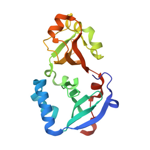 | |
UniProt & NIH Common Fund Data Resources | |||||
PHAROS: Q9ULC4 GTEx: ENSG00000232119 | |||||
Entity Groups | |||||
| Sequence Clusters | 30% Identity50% Identity70% Identity90% Identity95% Identity100% Identity | ||||
| UniProt Group | Q9ULC4 | ||||
Sequence AnnotationsExpand | |||||
Reference Sequence | |||||
| Length ( Å ) | Angle ( ˚ ) |
|---|---|
| a = 115.59 | α = 90 |
| b = 115.59 | β = 90 |
| c = 157.79 | γ = 120 |
| Software Name | Purpose |
|---|---|
| XSCALE | data scaling |
| SHELX | phasing |
| REFMAC | refinement |
| PDB_EXTRACT | data extraction |
| DENZO | data reduction |
| XDS | data reduction |
| SCALEPACK | data scaling |
| Coot | model building |
| MolProbity | model building |