3QO2 | pdb_00003qo2

Structural insights for MPP8 chromodomain interaction with histone H3 lysine 9


Experimental Data Snapshot

  • Method: X-RAY DIFFRACTION
  • Resolution: 2.49 Å
  • R-Value Free: 
    0.289 (Depositor), 0.283 (DCC) 
  • R-Value Work: 
    0.215 (Depositor), 0.209 (DCC) 
  • R-Value Observed: 
    0.218 (Depositor) 

Starting Model: experimental
View more details

wwPDB Validation 3D Report Full Report

Validation slider image for 3QO2

This is version 1.4 of the entry. See complete history

Literature

Structural insights for MPP8 chromodomain interaction with histone H3 lysine 9: potential effect of phosphorylation on methyl-lysine binding.

Chang, Y.Horton, J.R.Bedford, M.T.Zhang, X.Cheng, X.

(2011) J Mol Biology 408: 807-814

  • DOI: https://doi.org/10.1016/j.jmb.2011.03.018
  • Primary Citation Related Structures: 
    3QO2

  • PubMed Abstract: 

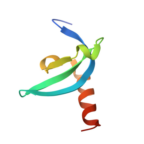
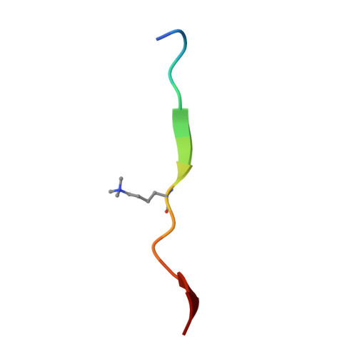
    M-phase phosphoprotein 8 (MPP8) harbors an N-terminal chromodomain and a C-terminal ankyrin repeat domain. MPP8, via its chromodomain, binds histone H3 peptide tri- or di-methylated at lysine 9 (H3K9me3/H3K9me2) in submicromolar affinity. We determined the crystal structure of MPP8 chromodomain in complex with H3K9me3 peptide. MPP8 interacts with at least six histone H3 residues from glutamine 5 to serine 10, enabling its ability to distinguish lysine-9-containing peptide (QTARKS) from that of lysine 27 (KAARKS), both sharing the ARKS sequence. A partial hydrophobic cage with three aromatic residues (Phe59, Trp80 and Tyr83) and one aspartate (Asp87) encloses the methylated lysine 9. MPP8 has been reported to be phosphorylated in vivo, including the cage residue Tyr83 and the succeeding Thr84 and Ser85. Modeling a phosphate group onto the side-chain hydroxyl oxygen of Tyr83 suggests that the negatively charged phosphate group could enhance the binding of positively charged methyl-lysine or create a regulatory signal by allowing or inhibiting binding of other protein(s).


  • Organizational Affiliation
    • Department of Biochemistry, Emory University School of Medicine, Atlanta, GA 30322, USA.

Macromolecule Content 

  • Total Structure Weight: 37.07 kDa 
  • Atom Count: 2,460 
  • Modeled Residue Count: 295 
  • Deposited Residue Count: 316 
  • Unique protein chains: 2

Macromolecules

Find similar proteins by:|  3D Structure
Entity ID: 1
MoleculeChains  Sequence LengthOrganismDetailsImage
M-phase phosphoprotein 8A,
C [auth B],
E [auth C],
G [auth D]
64Homo sapiensMutation(s): 0 
Gene Names: MPHOSPH8MPP8
UniProt & NIH Common Fund Data Resources
Find proteins for Q99549 (Homo sapiens)
Explore Q99549 
Go to UniProtKB:  Q99549
PHAROS:  Q99549
GTEx:  ENSG00000196199 
Entity Groups
Sequence Clusters30% Identity50% Identity70% Identity90% Identity95% Identity100% Identity
UniProt GroupQ99549
Sequence Annotations
Expand
Reference Sequence
Find similar proteins by:  Sequence   |   3D Structure  
Entity ID: 2
MoleculeChains  Sequence LengthOrganismDetailsImage
Histone H3 peptideB [auth P],
D [auth Q],
F [auth R],
H [auth S]
15synthetic constructMutation(s): 0 
UniProt & NIH Common Fund Data Resources
Find proteins for P68431 (Homo sapiens)
Explore P68431 
Go to UniProtKB:  P68431
Entity Groups
Sequence Clusters30% Identity50% Identity70% Identity90% Identity95% Identity100% Identity
UniProt GroupP68431
Sequence Annotations
Expand
Reference Sequence

Small Molecules

Experimental Data & Validation

Experimental Data

  • Method: X-RAY DIFFRACTION
  • Resolution: 2.49 Å
  • R-Value Free:  0.289 (Depositor), 0.283 (DCC) 
  • R-Value Work:  0.215 (Depositor), 0.209 (DCC) 
  • R-Value Observed: 0.218 (Depositor) 
Space Group: P 43 21 2
Unit Cell:
Length ( Å )Angle ( ˚ )
a = 53.461α = 90
b = 53.461β = 90
c = 238.792γ = 90
Software Package:
Software NamePurpose
HKL-2000data collection
PHENIXmodel building
PHENIXrefinement
HKL-2000data reduction
HKL-2000data scaling
PHENIXphasing

Structure Validation

View Full Validation Report


Entry History 

Deposition Data

Revision History  (Full details and data files)

  • Version 1.0: 2011-04-06
    Type: Initial release
  • Version 1.1: 2011-07-13
    Changes: Version format compliance
  • Version 1.2: 2012-02-29
    Changes: Database references
  • Version 1.3: 2017-11-08
    Changes: Refinement description
  • Version 1.4: 2023-09-13
    Changes: Data collection, Database references, Derived calculations, Refinement description