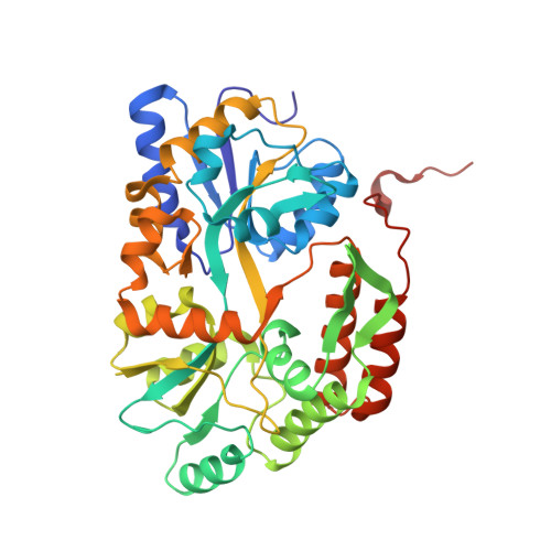
3Q28

Cyrstal structure of human alpha-synuclein (58-79) fused to maltose binding protein (MBP)


Experimental Data Snapshot

  • Method: X-RAY DIFFRACTION
  • Resolution: 1.60 Å
  • R-Value Free: 0.173 
  • R-Value Work: 0.152 
  • R-Value Observed: 0.153 

Starting Model: experimental
View more details

wwPDB Validation   3D Report Full Report


This is version 2.1 of the entry. See complete history


Literature

Structures of segments of alpha-synuclein fused to maltose-binding protein suggest intermediate states during amyloid formation

Zhao, M.Cascio, D.Sawaya, M.R.Eisenberg, D.

(2011) Protein Sci 20: 996-1004

  • DOI: https://doi.org/10.1002/pro.630
  • Primary Citation of Related Structures:  
    3Q25, 3Q26, 3Q27, 3Q28, 3Q29

  • PubMed Abstract: 

    Aggregates of the protein α-synuclein are the main component of Lewy bodies, the hallmark of Parkinson's disease. α-Synuclein aggregates are also found in many human neurodegenerative diseases known as synucleinopathies. In vivo, α-synuclein associates with membranes and adopts α-helical conformations. The details of how α-synuclein converts from the functional native state to amyloid aggregates remain unknown. In this study, we use maltose-binding protein (MBP) as a carrier to crystallize segments of α-synuclein. From crystal structures of fusions between MBP and four segments of α-synuclein, we have been able to trace a virtual model of the first 72 residues of α-synuclein. Instead of a mostly α-helical conformation observed in the lipid environment, our crystal structures show α-helices only at residues 1-13 and 20-34. The remaining segments are extended loops or coils. All of the predicted fiber-forming segments based on the 3D profile method are in extended conformations. We further show that the MBP fusion proteins with fiber-forming segments from α-synuclein can also form fiber-like nano-crystals or amyloid-like fibrils. Our structures suggest intermediate states during amyloid formation of α-synuclein.


  • Organizational Affiliation

    Department of Biological Chemistry, David Geffen School of Medicine, University of California, Los Angeles, California 90095, USA.


Macromolecules
Find similar proteins by:  (by identity cutoff)  |  3D Structure
Entity ID: 1
MoleculeChains Sequence LengthOrganismDetailsImage
Maltose-binding periplasmic protein/alpha-synuclein chimeric protein393Escherichia coliHomo sapiens
This entity is chimeric
Mutation(s): 0 
Gene Names: SNCA
UniProt & NIH Common Fund Data Resources
Find proteins for P0AEX9 (Escherichia coli (strain K12))
Explore P0AEX9 
Go to UniProtKB:  P0AEX9
Find proteins for P37840 (Homo sapiens)
Explore P37840 
Go to UniProtKB:  P37840
PHAROS:  P37840
GTEx:  ENSG00000145335 
Entity Groups  
Sequence Clusters30% Identity50% Identity70% Identity90% Identity95% Identity100% Identity
UniProt GroupsP37840P0AEX9
Sequence Annotations
Expand
  • Reference Sequence
Oligosaccharides

Help

Entity ID: 2
MoleculeChains Length2D Diagram Glycosylation3D Interactions
alpha-D-glucopyranose-(1-4)-alpha-D-glucopyranose
B
2N/A
Glycosylation Resources
GlyTouCan:  G07411ON
GlyCosmos:  G07411ON
Biologically Interesting Molecules (External Reference) 1 Unique
Experimental Data & Validation

Experimental Data

  • Method: X-RAY DIFFRACTION
  • Resolution: 1.60 Å
  • R-Value Free: 0.173 
  • R-Value Work: 0.152 
  • R-Value Observed: 0.153 
  • Space Group: P 1 21 1
Unit Cell:
Length ( Å )Angle ( ˚ )
a = 57.54α = 90
b = 48.57β = 94.23
c = 57.85γ = 90
Software Package:
Software NamePurpose
XSCALEdata scaling
PHASERphasing
PHENIXrefinement
PDB_EXTRACTdata extraction
XDSdata reduction

Structure Validation

View Full Validation Report


Entry History 

Deposition Data

Revision History  (Full details and data files)

  • Version 1.0: 2011-06-01
    Type: Initial release
  • Version 1.1: 2011-07-13
    Changes: Version format compliance
  • Version 1.2: 2017-08-02
    Changes: Refinement description, Source and taxonomy
  • Version 1.3: 2017-10-11
    Changes: Data collection
  • Version 2.0: 2020-07-29
    Type: Remediation
    Reason: Carbohydrate remediation
    Changes: Atomic model, Data collection, Database references, Derived calculations, Non-polymer description, Structure summary
  • Version 2.1: 2023-09-13
    Changes: Data collection, Database references, Refinement description, Structure summary